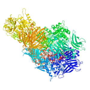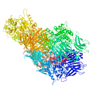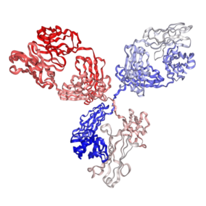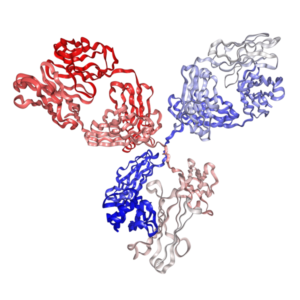
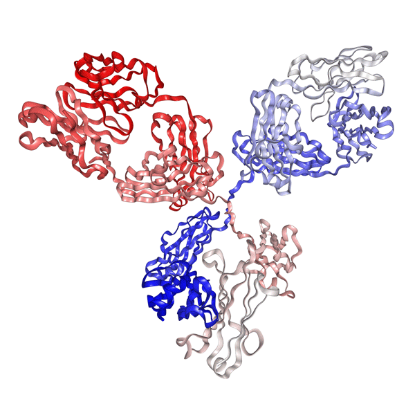

Anti-mouse PD-1 Antibody (CD279, RMP1-14.1) | PA007162.r2a
$150.00 – $700.00
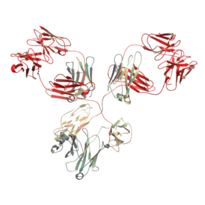
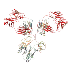
Our recombinant anti-mouse PD-1 antibody (Clone RMP1-14.1, Rat IgG2a, kappa) features the same variable region sequence as the original rat anti-mouse PD-1 monoclonal antibody (Clone RMP1-14). Recombinantly expressed in mammalian cells, this in vivo grade antibody is the most popular choice for immune checkpoint blockade studies in MC38 and CT26 mouse tumor models.
- Target & Clone: Mouse PD-1 / CD279 (Clone RMP1-14.1)
- Isotype: Rat IgG2a, kappa
- Binding Affinity: High affinity to mouse PD-1 protein (KD < 2 nM)
- Applications: In vivo PD-1 neutralization, T-cell reactivation, syngeneic tumor models
- Matching Control: Recombinant Rat IgG2a Isotype Control Antibody is available.
- Details & Specifications
- References
- Other Formats
- Ushelf Advisor
| Catalog No. | PA007162.r2a |
|---|---|
| Product Name | In Vivo Grade Recombinant Anti-mouse PD-1 Monoclonal Antibody (Clone RMP1-14.1), Rat IgG2a Kappa |
| Data Sheet | 📄 Download Product Data Sheet (PDF) |
| Supplier Name | Syd Labs, Inc. |
| Brand Name | Syd Labs |
| Synonyms | Programmed Cell Death Protein 1, CD279 antibody |
| Summary | The Anti-mouse PD-1 Antibody (RMP1-14.1, Rat IgG2a Kappa ) was produced in mammalian cells. |
| Clone | RMP1-14.1, the same variable region and constant region sequences as the rat anti-mouse PD-1 monoclonal antibody (clone number: RMP1-14) |
| Isotype | Rat IgG2a, kappa |
| Applications | immunohistochemistry (IHC), Flow Cytometry (FC), and various in vitro and in vivo functional assays. |
| Immunogen | The original rat hybridoma (clone name: RMP1-14) was generated by immunizing Sprague Dawley rats with mouse PD-1-transfected BHK cells and using a P3U1 myeloma as the fusion partner. |
| Form Of Antibody | 0.2 μM filtered solution of 1x PBS. |
| Endotoxin | Less than 1 EU/mg of protein as determined by LAL method. |
| Purity | >95% by SDS-PAGE under reducing conditions. |
| Shipping | The In Vivo Grade Recombinant Anti-mouse PD-1 Antibody (RMP1-14.1) is shipped with ice pack. Upon receipt, store it immediately at the temperature recommended below. |
| Stability & Storage | Use a manual defrost freezer and avoid repeated freeze-thaw cycles. 1 month from date of receipt, 2 to 8°C as supplied. 3 months from date of receipt, -20°C to -70°C as supplied. |
| RRID | RRID: AB_3697971 |
| Note | Recombinant mouse anti-mouse PD 1 / CD279 monoclonal antibodies, whose variable region sequences are murined from the rat anti-mouse PD-1 monoclonal antibody (clone number: RMP1-14), are produced from mammalian cells. The recombinant rat and chimeric mouse versions of the RMP1-14 antibody are also available. |
| Order Offline | Phone: 1-617-401-8149 Fax: 1-617-606-5022 Email: message@sydlabs.com Or leave a message with a formal purchase order (PO) Or credit card. |
Description
Recombinant Anti-mouse PD-1 (CD279) Antibody – Clone RMP1-14.1: The Precision Evolution of RMP1-14
The Recombinant Anti-mouse PD-1 Monoclonal Antibody (Clone RMP1-14.1) represents the next generation of checkpoint inhibitors for immunotherapy research. As a sequence-defined, recombinant version of the classic RMP1-14 clone, RMP1-14.1 is engineered to eliminate the inherent variability of hybridoma-derived antibodies, providing researchers with 100% batch-to-batch consistency and superior molecular integrity.
Targeting the CD279 (Programmed Death-1) receptor, this antibody is specifically optimized for in vivo PD-1 blockade in syngeneic mouse models. Unlike traditional RMP1-14, our recombinant RMP1-14.1 is produced in a specialized mammalian expression system, ensuring it is animal-free, ultra-pure, and carries the precise in vivo functional grade required for high-impact preclinical studies.
Why RMP1-14.1 is the New Gold Standard:
-
Engineered for Reproducibility: Being sequence-defined, RMP1-14.1 eliminates the risk of “gene loss” often seen in hybridoma cell lines, guaranteeing identical performance across every vial.
-
Validated for Tumor Models: Extensively tested in MC38, CT26, and B16-F10 syngeneic models to ensure potent neutralization of the PD-1/PD-L1 axis.
-
Advanced Fc-Engineering: Available in multiple formats, including Rat IgG2a, Mouse IgG1/IgG2a, and Fc-silent (LALAPG) mutations to minimize non-specific Fc-receptor binding during sensitive T-cell exhaustion studies.
Unlike traditional RMP1-14 hybridomas which are prone to genetic drift and lot-to-lot variation, Syd Labs’ Recombinant RMP1-14.1 is sequence-defined. This ensures that the antibody you use in 2026 will perform exactly the same as the batch you used five years ago. For researchers performing T-cell activation assays alongside checkpoint inhibition, this antibody is the ideal companion to Syd Labs’ Recombinant OKT3 (Anti-human CD3) and other effector-cell modulators.
Citations: This recombinant clone is gaining rapid traction as a high-reproducibility alternative to hybridoma-derived BE0146.
References about Anti-mouse PD-1 (CD279, RMP1-14.1) MAb,please click: anti-mouse PD-1 monoclonal antibody (clone RMP1-14) referenced literature.
References of Anti-Mouse PD-1 Monoclonal Antibody (Clone: RMP1-14.1)
1. Spermidine potentiates anti-tumor immune responses and immunotherapy sensitivity in breast cancer
Xinyu Yang, et al. J Cancer 2025. doi: 10.7150/jca.113235
“In vivo spermidine supplementing experiment…To establish a subcutaneous tumor-bearing mouse model …… Docetaxel (Sanofi Mature IP) was administered intraperitoneally at 10 mg/kg one week after tumor inoculation in mice, followed by a single intraperitoneal injection of PD-1 antibody (SYD, PA007162) at a dosage of 10 mg/kg …… The Institutional Animal Care and Use Committee at Sun Yat-Sen University granted approval for animal experiments.”
2. PD-L1/PD-1 checkpoint pathway regulates hippocampal neuronal excitability and learning and memory behavior
Junli Zhao, et al. Neuron 2023. doi: 10.1016/j.neuron.2023.07.008
“Programmed death protein 1 (PD-1) and its ligand PD-L1 constitute an immune checkpoint pathway. We report that neuronal PD-1 signaling regulates learning/memory in health and disease. Intraventricular administration of anti-mouse PD-1 monoclonal antibody (RMP1-14) potentiated learning and memory.”
3. PD-L1 signaling selectively regulates T cell lymphatic transendothelial migration
Wenji Piao, et al. Nat Commun 2022. doi: 10.1038/s41467-022-29761-9
“Programmed death-1 (PD-1) and its ligand PD-L1 are checkpoint molecules which regulate immune responses. Antibody blockade of Treg PD-1, Teff CD80 (the alternative ligand for PD-L1), or LEC PD-L1 impairs Treg or Teff migration in vitro and in vivo. PD-1/PD-L1 signals through PI3K/Akt and ERK to regulate zipper junctional VE-cadherin, and through NFκB-p65 to up-regulate VCAM-1 expression on LECs.”
4. Macrophages Impair TLR9 Agonist Antitumor Activity through Interacting with the Anti-PD-1 Antibody Fc Domain
Simone Camelliti, et al. Cancers (Basel) 2021. doi: 10.3390/cancers13164081
“We evaluated the contribution of macrophages to the effect of combinatorial immunotherapeutic treatments based on TLR9 stimulation (with CpG-ODNs) and PD-1 blockade in an ovarian cancer preclinical model. We observed a strong reduction in the antitumor efficacy of a TLR9 agonist upon anti-PD-1 antibody administration. Specifically, we found that TLR9-stimulated macrophages, through interacting with the fragment crystallizable (Fc) domain of the anti-PD-1 antibody, acquire an immunoregulatory phenotype leading to dampening of CpG-ODN antitumor effect.”
5. Prophylactic IL-23 blockade uncouples efficacy and toxicity in dual CTLA-4 and PD-1 immunotherapy
Mingyi Ju, et al. J Immunother Cancer 2024. doi: 10.1136/jitc-2024-009144
“The onset of immune-related adverse events (irAEs) might serve as a clinical biomarker to predict a favorable therapeutic response to immune checkpoint inhibitors (ICIs). Moreover, in the PD-1 monotherapy cohort, patients with irAEs tended to achieve higher response rates than those lacking toxicity. Our findings fill the gap in the previous evidence that there was no correlation between irAEs and ICIs response in patients treated with anti-CTLA-4 therapy.”
6. Angiogenic inhibitor pre‐administration improves the therapeutic effects of immunotherapy
Ying Han, et al. Cancer Sci 2023. doi: 10.1111/cas.15783
“Angiogenic inhibitors combined with anti-PD-1 blockade has become a standard choice for multiple advanced malignancies. We demonstrated that pre-treatment with anti-angiogenic agents improves tumor vascular normalization. This optimal pre-administration schedule enhances the subsequent therapeutic efficacy of anti-mouse PD-1 antibody treatment in syngeneic models.”
7. Sources of inter-individual variability leading to significant changes in anti-PD-1 and anti-PD-L1 efficacy identified in mouse tumor models using a QSP framework
Chetan Kulkarni, et al. CPT Pharmacometrics Syst Pharmacol 2022. doi: 10.1002/psp4.12879
“Substantial inter-individual variability in response to immune checkpoint inhibition is observed clinically and in mouse models. We applied a quantitative systems pharmacology (QSP) model to investigate physiological drivers of variability. Our simulations identify critical parameters regulating anti-PD-1 efficacy and receptor occupancy profiles in murine syngeneic cohorts.”
8. Targeting PD-L2/RGMb overcomes microbiome-related immunotherapy resistance
Jong-Chan Park, et al. Nature 2023. doi: 10.1038/s41586-023-06103-x
“The gut microbiota significantly influences the response of tumors to immune checkpoint blockades. We identify that a subset of commensal microbes impairs anti-PD-1 therapeutic outcomes through distinct pathways. Concurrent blockade targeting the PD-L2/RGMb axis effectively overcomes this resistance and rescues anti-PD-1 treatment efficacy.”
9. Targeted delivery of a PD-1-blocking scFv by CAR-T cells enhances anti-tumor efficacy in vivo
Stephen J. Price, et al. Nat Biotechnol 2018. doi: 10.1038/nbt.4194
“Combining CAR-T cells with immune checkpoint inhibitors represents an attractive strategy to counter immunosuppressive environments. We engineered CAR-T cells to locally secrete an anti-PD-1 single-chain variable fragment (scFv). This localized action achieved superior therapeutic antitumor clearance compared to systemic combination with standard anti-PD-1 antibodies.”
10. Antibody-mediated depletion of programmed death 1-positive (PD-1+) cells
Takahiro Shimizu, et al. Immunology 2023. doi: 10.1111/imm.13702
“Anti-PD-1 antibodies are typically used to block inhibitory signals in dysfunctional T cells. Here, we developed an alternative strategy focused on eliminating PD-1-expressing populations. Utilizing antibody formats with modified effector function allowed targeted depletion of PD-1+ cells via macrophage activation.”
11. Synergistic anticancer activity of a novel oral chemotherapeutic agent containing trifluridine and tipiracil in combination with anti-PD-1 blockade in microsatellite stable-type murine colorectal cancer cells
Tatsuro Suenaga, et al. Oncotarget 2017. doi: 10.18632/oncotarget.20455
“Microsatellite stable colorectal cancer displays limited response to conventional anti-PD-1 blockades. We evaluated the combined efficacy of oral trifluridine/tipiracil and systemic anti-PD-1 monoclonal antibodies. This combination significantly enhanced tumor growth inhibition and CD8+ T-cell infiltration into syngeneic lesions.”
12. Type I MET inhibitors cooperate with PD-1 blockade to promote rejection of hepatocellular carcinoma
Elena Martinez, et al. J Hepatol 2024. doi: 10.1016/j.jhep.2024.05.011
“Hepatocellular carcinoma often creates an immune-excluded microenvironment resisting single-agent treatments. We showed that selective type I MET inhibition reverses immune exclusion patterns. Combining this targeted approach with anti-PD-1 antibody interventions triggers complete rejection in a high percentage of murine models.”
13. Dual targeting of RANKL and PD‐1 with a bispecific antibody improves anti‐tumor immunity
David A. Khan, et al. Clin Transl Immunology 2019. doi: 10.1002/cti2.1075
“Targeting RANKL alters the osteoclast niche and can modulate immunosuppressive myeloid components. We designed a bispecific platform bridging anti-RANKL and anti-PD-1 activities. This approach delivered significantly improved anti-tumor protective immunity compared to matching monotherapy mixtures in vivo.”
14. Lymph node and tumor-associated PD-L1+ macrophages antagonize dendritic cell vaccines by suppressing CD8+ T cells
Nathalie M. Laureano, et al. J Clin Invest 2024. doi: 10.1172/JCI172031
“Dendritic cell vaccinations frequently exhibit sub-optimal clinical outcomes due to local active suppression. We demonstrate that PD-L1-expressing macrophages in regional nodes directly suppress functional vaccine responses. Administering anti-PD-1 therapeutic agents effectively counteracts this macrophage-mediated antagonism.”
15. Decoupling FcRn and tumor contributions to elevated immune checkpoint inhibitor clearance in cancer cachexia
Alexander R. S. Johnson, et al. J Cachexia Sarcopenia Muscle 2023. doi: 10.1002/jcsm.13340
“Cancer cachexia accelerates the clearance of multiple monoclonal therapeutic antibodies, reducing exposure. We assessed the specific contributions of target burden and FcRn-mediated recycling dynamics. Our findings outline that cachexia altered antibody elimination tracks independently of typical target receptor distributions.”
16. Diphtheria toxin‐derived, anti‐PD‐1 immunotoxin, a potent and practical tool to selectively deplete PD‐1+ cells
Yumi Matsuoka, et al. Immunity 2023. doi: 10.1016/j.immuni.2023.07.014
“Investigating the roles of chronic PD-1+ populations requires reliable depletion methods. We engineered a diphtheria toxin-based immunotoxin architecture targeting mouse PD-1 structures. This molecule mediates rapid, highly specific systemic elimination of active PD-1-positive subgroups inside immunocompetent strains.”
17. Targeting HIF-1α abrogates PD-L1–mediated immune evasion in tumor microenvironment but promotes tolerance in normal tissues
Marcus O. Scharping, et al. J Clin Invest 2022. doi: 10.1172/JCI156020
“Hypoxia-inducible factor 1α (HIF-1α) acts as a critical metabolic sensor regulating target expression. We show that conditional ablation of metabolic pathways selectively limits immune evasion mechanisms. Combining metabolic modulation with anti-PD-1 blockade achieves robust therapeutic responses while maintaining peripheral organ safety boundaries.”
18. Distinct antibody clones detect PD-1 checkpoint expression and block PD-L1 interactions on live murine melanoma cells
Christopher H. Smith, et al. PLoS One 2022. doi: 10.1371/journal.pone.0271501
“Accurate characterization of receptor blockades requires precise tracking of clone specificities. We systematically evaluated multiple commercial and recombinant monoclonal antibody lines on active melanoma targets. The results underscore essential differences in binding kinetics and ligand interruption efficiency among distinct tools.”
19. Immune receptor inhibition through enforced phosphatase recruitment
Rachel N. Vance, et al. Science 2021. doi: 10.1126/science.abe7542
“Inhibitory pathways typically signal via localized phosphatase clustering to disrupt downstream activation complexes. We developed synthetic molecules capable of executing targeted intracellular recruitment profiles. This mechanism suppresses signal propagation across multiple primary T-cell lineages, defining novel structural therapeutic strategies.”
20. Immune tolerance against infused FVIII in hemophilia A is mediated by PD-L1+ Tregs
Thomas M. Miller, et al. Blood 2022. doi: 10.1182/blood.2022016540
“Inhibitor development against replacement factor VIII (FVIII) represents a critical clinical challenge in hemophilia management. We demonstrated that establishing functional peripheral tolerance relies heavily on specific regulatory populations. Disrupting the pathway via anti-PD-1 administration completely breaks factor protection in experimental models.”
21. Myeloid Antigen-Presenting Cell Niches Sustain Antitumor T Cells and License PD-1 Blockade via CD28 Costimulation
Brian C. Gubin, et al. Cell 2022. doi: 10.1016/j.cell.2022.01.018
“Intratumoral T-cell expansion and maintenance require sustained supportive micro-environments. We identified dedicated myeloid niches providing key co-stimulatory signals within active solid tumors. Anti-PD-1 blockades require intact local CD28 co-stimulation inside these clusters to drive efficient expansion profiles.”
For more technical references or data sheets regarding the Recombinant Anti-Mouse PD-1 Monoclonal Antibody (Clone: RMP1-14.1) formats, including options from Syd Labs, please contact our scientific support team at message@sydlabs.com.
Related Recombinant IgG Reference Antibodies:
Recombinant Mouse IgG1 Isotype Control Antibody and Mutants, In vivo Grade
Recombinant Mouse IgG2a Isotype Control Antibody and Mutants, In vivo Grade
Recombinant Mouse IgG2c Isotype Control Antibody and Mutants, In vivo Grade
Recombinant Rat IgG2a Isotype Control Antibody, In vivo Grade
Syd Labs provides the following anti-mouse PD-L1 / PD-1 antibodies:
Recombinant anti-mouse PD1 antibodies (Clone 29F.1A12.1), In vivo grade
Recombinant anti-mouse PD-1 antibodies (Clone RMP1-14.1), In vivo grade
Recombinant anti-mouse PD-L1 antibodies (Clone 10F.9G2.1), In vivo grade
Recombinant anti-mouse PD-1 / PD-1 bispecific antibodies (Clone RMP1-14.1 / 29F.1A12.1), In vivo grade
Recombinant anti-mouse PD-1 / PD-1 bispecific antibodies (Clone 29F.1A12.1 / RMP1-14.1), In vivo grade
Recombinant anti-mouse PD-1 / PD-L1 bispecific antibodies (Clone RMP1-14.1 / 10F.9G2.1), In vivo grade
Recombinant anti-mouse PD-L1 / PD-1 bispecific antibodies (Clone 10F.9G2.1 / RMP1-14.1), In vivo grade
Recombinant anti-mouse PD-1 / PD-L1 bispecific antibodies (Clone 29F.1A12.1 / 10F.9G2.1), In vivo grade
Recombinant anti-mouse PD-L1 / PD-1 bispecific antibodies (Clone 10F.9G2.1 / 29F.1A12.1), In vivo grade
Other Formats
| Product Name | Catalog No. | Isotype | Host / Reactivity | Applications |
|---|---|---|---|---|
| In Vivo Grade Recombinant Anti-mouse PD-1 Monoclonal Antibody (Clone RMP1-14.1), Mouse IgG2a Kappa | PA007162.m2a | Mouse IgG2a, kappa | Mouse / Mouse | IHC, FC, in vitro and in vivo functional assays |
| In Vivo Grade Recombinant Anti-mouse PD-1 Monoclonal Antibody (Clone RMP1-14.1), Mouse IgG1 Kappa | PA007162.m1 | Mouse IgG1, kappa | Mouse / Mouse | IHC, FC, in vitro and in vivo functional assays |
| In Vivo Grade Recombinant Anti-mouse PD-1 Monoclonal Antibody (Clone RMP1-14.1), Mouse IgG2c-L234A L235A P329G (LALAPG) Kappa | PA007162.m2cLA | Mouse IgG2c LALAPG, kappa Fc-Silenced | Mouse / Mouse | IHC, FC, in vitro and in vivo functional assays, Fc-silenced neutralization |
| In Vivo Grade Recombinant Anti-mouse PD-1 Monoclonal Antibody (Clone RMP1-14.1), Mouse IgG2a-L234A L235A P329G (LALAPG) Kappa | PA007162.m2aLA | Mouse IgG2a LALAPG, kappa Fc-Silenced | Mouse / Mouse | IHC, FC, in vitro and in vivo functional assays, Fc-silenced neutralization |
| Anti-mouse PD-1 (RMP1-14.1) Antibody , Mouse IgG1-D265A Kappa | PA007162.m1DA | Mouse IgG1-D265A, kappa Fc-Silenced | Mouse / Mouse | IHC, FC, in vitro and in vivo functional assays, Fc-silent neutralization |
| In Vivo Grade Recombinant Anti-mouse PD-1 Monoclonal Antibody (Clone RMP1-14.1), Rabbit IgG | PA007162.rt | Rabbit IgG | Rabbit / Mouse | IHC, FC, in vitro and in vivo functional assays |
Ushelf Advisor
Powered by AI: AI is experimental and still learning how to provide the best assistance. It may occasionally generate incorrect or incomplete responses. Please do not rely solely on its recommendations when making purchasing decisions or designing experiments.
Q: What is the recommended in vivo dosage for anti-mouse PD-1 antibody in tumor models?
There is no single universally “recommended” dosage for anti-mouse PD-1 antibodies (e.g., clone RMP1-14, the most widely used) in syngeneic tumor models, as it varies based on factors like tumor type (e.g., MC38, B16, CT26), mouse strain (commonly C57BL/6), timing relative to tumor inoculation, combination therapies, and desired effect (pure blockade vs. partial depletion).
However, based on extensive use in published studies and other supplier guidelines, the most common regimens are:
Dose per injection: 200–500 μg per mouse (typically ~10–25 mg/kg for a 20–25 g mouse). The lower end (200–250 μg or ~10 mg/kg) is most frequently reported for effective PD-1 blockade with minimal toxicity in responsive models.
Frequency and schedule: Every 3–5 days, often for 3–6 doses total. A classic protocol is injections on days 3, 6, 9 (or similar) post-tumor implantation, with additional doses as needed.
Route of administration: Intraperitoneal (i.p.) injection is standard.
Q: Which anti-mouse PD1 antibody works the best?
There are several anti-mouse PD1 antibodies available in the market: Clones RMP1-14, 29F.1A12, and J43. All three of these antibodies are commonly used to block PD-1 signaling in vivo in murine tumor models and other mouse models. These three clones all have extensive multi-year publication records supporting them. The RMP1-14 antibody has been reported to block the binding of PD-1 to its ligands (B7-H1 and B7-DC) and to inhibit T cell proliferation and cytokine production costimulated by macrophages (but not by dendritic cells and B cells). (by Syd Labs) Syd Labs offers anti-mouse PD-1 monoclonal antibodies based on the sequences of clones RMP1-14 and 29F.1A12. Syd Labs provides in vivo grade recombinant antibodies including engineered antibodies for the clones RMP1-14 and 29F.1A12. Even though mouse and rat are close, rat antibodies may still induce immunogenecity in mice. Antibodies with murinized variable regions and mouse constant regions behave like humanized antibody drugs in animal models using mice. In addition, the mouse IgG2c antibody is produced in certain inbred strains such as C57BL/6, C57BL/10, SJL, and NOD, which does not express the mouse IgG2a antibody; the mouse IgG2a antibody is produced in other inbred strains such as BALB/c and Swiss Webster mice, which does not express the mouse IgG2c antibody. If one uses the C57BL/6 mouse strain for animal model research, it is better to use the IgG2c antibodies rather than the IgG2a antibodies. The format with the Fc silenced, Fc silent, or Fcs with silenced effector function, such as LALAPG mutation, is the most popular for anti-mouse PD1 antibodies (clones RMP1-14 and 29F.1A12) and anti-mouse PD-L1 antibodies (clone 10F.9G2).
Q: Do you produce any recombinant Fc-silenced RMP1-14 antibody?
Sure, we provide various recombinant Fc silent RMP1-14 antibodies, such as mIgG2c LALAPG, mIgG2a LALAPG, and mIgG1 D265A. We also provide custom recombinant antibody production service to produce other engineered versions of recombinant RMP1-14 antibodies. We have a promotion program running: We provide 1 mg PA007162.m2cLA (In Vivo Grade Recombinant Anti-mouse PD-1 Mouse IgG2c-LALAPG Kappa Monoclonal Antibody (Clone RMP1-14.1)) for free in exchange of results. Please contact us to know more about the free RMP1-14 antibody.
Q: What is the difference among PA007162.r2a, PA007162.m2cLA, and PA007162.mm2cLA?
PA007162.r2a is the recombinant anti-mouse PD-1 monoclonal antibody (rat IgG2a kappa, clone RMP1-14.1) produced in CHO cells or HEK293 cells if needed. It has the same variable region and constant region sequences as the rat anti-mouse PD-1 monoclonal antibody from the hybridoma clone of RMP1-14. Rat antibodies may cause high immuogenicity in mice; thus, at least recombinant antibodies with mouse antibody constant regions should be used to replace the rat antibody constant regions. PA007162.m2cLA is the recombinant anti-mouse PD-1 antibody (clone RMP1-14.1) whose constant regions are mouse IgG2c LALAPG kappa. We further murinize the antibody variable region sequences of PA007162.m2cLA to produce PA007162.mm2cLA.
You may also like…
-
 Select optionsQuick View
 -
 Select optionsQuick View
 -
 Select optionsQuick View
 -
 Select optionsQuick View
 -
 Select optionsQuick View
 -
 Select optionsQuick View
 -
 Select optionsQuick ViewAnti-Mouse Antibodies (In Vivo), Antibodies
Anti-mouse PD-1 (CD279)-InVivo | PA007162.m2a
$150.00 – $1,100.00
![Anti-Mouse PD-1 (CD279) [RMP1-14] In Vivo Antibody | PA007162.rt Ushelf](https://www.ushelf.com/wp-content/uploads/2025/04/Anti-Mouse-PD-1-CD279-RMP1-14-In-Vivo-Antibody-300x300.png)