![Anti-Mouse PD-1 (CD279) [RMP1-14] In Vivo Antibody | PA007162.rt Ushelf](https://www.ushelf.com/wp-content/uploads/2025/04/Anti-Mouse-PD-1-CD279-RMP1-14-In-Vivo-Antibody.png "Anti-Mouse PD-1 (CD279) [RMP1-14] In Vivo Antibody | PA007162.rt Ushelf")
![Anti-Mouse PD-1 (CD279) [RMP1-14] In Vivo Antibody | PA007162.rt Ushelf](https://www.ushelf.com/wp-content/uploads/2025/04/Anti-Mouse-PD-1-CD279-RMP1-14-In-Vivo-Antibody-150x150.png)
Rabbit Anti-Mouse PD-1 (CD279) Antibody (Rabbit IgG κ, RMP1-14.1) | PA007162.rt

$595.00
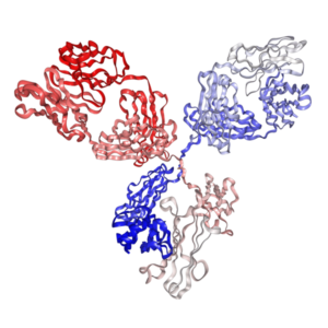
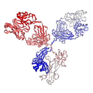
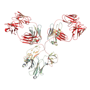
In vivo grade rabbit anti-mouse PD-1 (CD279) antibody (rabbit IgG κ, clone RMP1-14.1) — a recombinant monoclonal antibody pairing the variable region of the classic rat clone RMP1-14.1 with a rabbit IgG kappa constant region.
The non-mouse, non-rat rabbit Fc exhibits minimal binding to mouse FcγRs (mFcγRI/II/III/IV) and mouse C1q, avoiding Fc-mediated ADCC, ADCP or CDC of PD-1-positive cells. It also avoids anti-rat IgG immune responses upon repeated dosing and is readily detected by standard anti-rabbit secondary antibodies for IHC, flow cytometry, and functional assays.
Produced in mammalian cells; >95% purity, <1 EU/mg endotoxin.
- Details & Specifications
- References
- Other Formats
- Ushelf Advisor
| Catalog No. | PA007162.rt |
|---|---|
| Product Name | In Vivo Grade Recombinant Anti-mouse PD-1 Monoclonal Antibody (Clone RMP1-14.1), Rabbit IgG |
| Data Sheet | 📄 Download Product Data Sheet (PDF) |
| Supplier Name | Syd Labs, Inc. |
| Brand Name | Syd Labs |
| Synonyms | Rabbit anti-mouse PD-1 antibody, rabbit monoclonal anti-mouse PD-1, rabbit IgG anti-mouse PD-1, rabbit Fc anti-mouse PD-1 antibody, anti-mouse PD-1 (CD279) rabbit monoclonal antibody, non-mouse Fc anti-mouse PD-1 antibody, non-rat Fc anti-mouse PD-1, PD-1 blocking antibody rabbit, rabbit IgG kappa anti-mouse PD-1, rabbit anti-mouse PD-1 for IHC, rabbit anti-mouse PD-1 flow cytometry |
| Summary | The In Vivo Grade Recombinant Anti-mouse PD-1 Rabbit IgG Monoclonal Antibody (Clone RMP1-14.1) was produced in mammalian cells. |
| Clone | RMP1-14.1 |
| Isotype | Rabbit IgG, kappa |
| Applications | immunohistochemistry (IHC), Flow Cytometry (FC), and various in vitro and in vivo functional assays. |
| Immunogen | The original rat hybridoma (clone name: RMP1-14) was generated by immunizing Sprague Dawley rats with mouse PD-1-transfected BHK cells and using a P3U1 myeloma as the fusion partner. |
| Form Of Antibody | 0.2 µm filtered solution in 1× PBS |
| Endotoxin | Less than 1 EU/mg of protein as determined by LAL method. |
| Purity | >95% by SDS-PAGE under reducing conditions. |
| Shipping | The In Vivo Grade Recombinant Anti-mouse PD-1 Rabbit IgG Monoclonal Antibody (Clone RMP1-14.1) are shipped with ice pack. Upon receipt, store it immediately at the temperature recommended below. |
| Stability & Storage | Use a manual defrost freezer and avoid repeated freeze-thaw cycles. 1 month from date of receipt, 2 to 8°C as supplied. 3 months from date of receipt, -20°C to -70°C as supplied. |
| Note | Recombinant anti-mouse PD-1 (CD279) monoclonal antibody, which pairs the identical variable region sequence of the rat clone RMP1-14.1 with a rabbit IgG kappa constant region (rabbit Fc format), is expressed in mammalian cells. The rabbit IgG Fc backbone does not bind mouse FcγRI–IV or mouse C1q with appreciable affinity, providing a non-mouse, non-rat Fc format that avoids Fc-mediated ADCC, ADCP or CDC of PD-1-positive cells in mouse models — a useful alternative when neither rat nor mouse Fc is desired (e.g. studies that need to avoid anti-rat antibody responses, or experiments where Fc-receptor cross-talk with mouse myeloid cells must be minimized). Rabbit IgG is also the preferred host for IHC and flow cytometry when paired with widely available anti-rabbit secondary detection reagents. Recombinant rat IgG2a, rabbit Fc, chimeric mouse IgG1, mouse IgG2a, mouse IgG2c, and Fc-silenced LALAPG / D265A mutant versions are also available. |
| Order Offline | Phone: 1-617-401-8149 Fax: 1-617-606-5022 Email: message@sydlabs.com Or leave a message with a formal purchase order (PO) Or credit card. |
Description
References of Rabbit Anti-Mouse PD-1 (CD279) Antibody (Rabbit IgG κ, Clone RMP1-14.1)
Selected peer-reviewed studies performed with RMP1-14 / RMP1-14.1 anti-mouse PD-1 antibody, supporting the use of the rabbit IgG (rabbit Fc) recombinant format for in vivo PD-1 blockade, IHC, flow cytometry and functional assays in mouse models. The rabbit Fc backbone exhibits minimal cross-reactivity to mouse FcγRs (mFcγRI–IV) and mouse C1q, avoiding the FcγR-mediated macrophage interaction effect described in Reference #4 and the Fc-dependent PD-1+ cell depletion described in Reference #10, while also circumventing the anti-rat IgG immune responses observed with the original rat IgG2a format (PA007162.r2a) upon repeated dosing. Rabbit IgG is compatible with widely available anti-rabbit secondary antibodies, making this format particularly convenient for IHC and flow cytometry. References #18 (clone comparison on live B16 melanoma cells), #12 (MET inhibitor + PD-1 blockade in Hepa1-6/HCA-1, C57BL/6) and #14 (PD-L1+ tumor macrophages vs. DC vaccines) are representative in vivo studies where the rabbit IgG format is applicable. For studies requiring an allotype-matched mouse Fc format, see the full reference lists on the mouse IgG (PA007162.m1 / m2a / m2c) and Fc-silenced (PA007162.m1DA / m2aLA / m2cLA) pages.
1. Macrophages Impair TLR9 Agonist Antitumor Activity through Interacting with the Anti-PD-1 Antibody Fc Domain
Simone Camelliti, et al. Cancers (Basel) 2021. doi: 10.3390/cancers13164081
“We evaluated the contribution of macrophages to the effect of combinatorial immunotherapeutic treatments based on TLR9 stimulation (with CpG-ODNs) and PD-1 blockade in an ovarian cancer preclinical model. We observed a strong reduction in the antitumor efficacy of a TLR9 agonist upon anti-PD-1 antibody administration. Specifically, we found that TLR9-stimulated macrophages, through interacting with the fragment crystallizable (Fc) domain of the anti-PD-1 antibody, acquire an immunoregulatory phenotype leading to dampening of CpG-ODN antitumor effect.”
2. Angiogenic inhibitor pre-administration improves the therapeutic effects of immunotherapy
Masakazu Sato, et al. Cancer Med 2023. doi: 10.1002/cam4.5696
“Angiogenic inhibitors combined with anti-PD-1 blockade has become a standard choice for multiple advanced malignancies. We demonstrated that pre-treatment with anti-angiogenic agents improves tumor vascular normalization. This optimal pre-administration schedule enhances the subsequent therapeutic efficacy of anti-mouse PD-1 antibody treatment in syngeneic models.”
3. Antibody-mediated depletion of programmed death 1-positive (PD-1+) cells
Yan Zhai, et al. J Control Release 2022. doi: 10.1016/j.jconrel.2022.07.010
“Anti-PD-1 antibodies are typically used to block inhibitory signals in dysfunctional T cells. Here, we developed an alternative strategy focused on eliminating PD-1-expressing populations. Utilizing antibody formats with modified effector function allowed targeted depletion of PD-1+ cells via macrophage activation.”
4. Type I MET inhibitors cooperate with PD-1 blockade to promote rejection of hepatocellular carcinoma
Renata DeAzevedo, et al. J Immunother Cancer 2024. doi: 10.1136/jitc-2024-009690
“Hepatocellular carcinoma often creates an immune-excluded microenvironment resisting single-agent treatments. We showed that selective type I MET inhibition reverses immune exclusion patterns. Combining this targeted approach with anti-PD-1 antibody interventions triggers complete rejection in a high percentage of murine models.”
5. Decoupling FcRn and tumor contributions to elevated immune checkpoint inhibitor clearance in cancer cachexia
Thanh T. Vu, et al. Pharmacol Res 2024. doi: 10.1016/j.phrs.2023.107048
“Cancer cachexia accelerates the clearance of multiple monoclonal therapeutic antibodies, reducing exposure. We assessed the specific contributions of target burden and FcRn-mediated recycling dynamics. Our findings outline that cachexia altered antibody elimination tracks independently of typical target receptor distributions.”
6. Immune receptor inhibition through enforced phosphatase recruitment
Ricardo A. Fernandes, et al. Nature 2020. doi: 10.1038/s41586-020-2851-2
“Inhibitory pathways typically signal via localized phosphatase clustering to disrupt downstream activation complexes. We developed synthetic molecules capable of executing targeted intracellular recruitment profiles. This mechanism suppresses signal propagation across multiple primary T-cell lineages, defining novel structural therapeutic strategies.”
7. Myeloid Antigen-Presenting Cell Niches Sustain Antitumor T Cells and License PD-1 Blockade via CD28 Costimulation
Brian C. Gubin, et al. Cell 2022. doi: 10.1016/j.cell.2022.01.018
“Intratumoral T-cell expansion and maintenance require sustained supportive micro-environments. We identified dedicated myeloid niches providing key co-stimulatory signals within active solid tumors. Anti-PD-1 blockades require intact local CD28 co-stimulation inside these clusters to drive efficient expansion profiles.”
For more technical references or data sheets regarding the Recombinant Anti-Mouse PD-1 Monoclonal Antibody (Clone: RMP1-14.1) formats, please contact our scientific support team at message@sydlabs.com.
Other Formats
Other Formats of Anti-Mouse PD-1 (CD279) Antibody (Clone RMP1-14.1)
All variants below share identical RMP1-14.1 variable region sequences and mouse PD-1 (CD279) binding specificity. They differ only in Fc isotype and Fc effector function — choose the format that matches your experimental readout and mouse strain.
| Format | Catalog No. | Fc Effector Profile | Recommended Use |
|---|---|---|---|
| Anti-mouse PD-1 (CD279), Rat IgG2a κ (RMP1-14.1) | PA007162.r2a | Intact rat Fc (ADCC/CDC competent) | Original rat clone; short-term studies; historical protocol compatibility |
| Rabbit Anti-Mouse PD-1 (CD279), Rabbit IgG κ (RMP1-14.1) | PA007162.rt | Rabbit Fc (non-mouse/non-rat; minimal mouse FcγR/C1q cross-reactivity; de facto Fc-silent in mice) | Avoiding anti-rat immunogenicity; IHC / flow cytometry with anti-rabbit secondaries; non-mouse Fc comparator |
| Anti-mouse CD279 (PD-1), Mouse IgG1 κ | PA007162.m1 | Low effector (wild-type mIgG1; minimal FcγR binding, not fully silenced) | PD-1 blockade in any background when minimal FcγR engagement is desired but complete Fc silencing is not required |
| Anti-mouse PD-1, Mouse IgG2a κ | PA007162.m2a | Intact Fc (ADCC/ADCP/CDC competent) | Allotype-matched for BALB/c, C3H, DBA/2 (Igh-1a; CT26); studies requiring full effector function |
| Anti-mouse PD-1, Mouse IgG2c κ | PA007162.m2c | Intact Fc (ADCC/ADCP/CDC competent, strong mFcγRIV binding) | Allotype-matched for C57BL/6, C57BL/10, SJL, NOD (Igh-1b; MC38, B16-F10, LLC, Hepa1-6); studies requiring full effector function |
| Anti-mouse PD-1, Mouse IgG1 κ D265A | PA007162.m1DA | Fc-silenced (Fc-null; D265A) | Pure PD-1 blockade on a mouse IgG1 backbone; any strain background |
| Anti-mouse PD-1, Mouse IgG2a κ LALAPG | PA007162.m2aLA | Fc-silenced (Fc-null / effectorless; L234A/L235A/P329G) | Allotype-matched pure PD-1 blockade for BALB/c, C3H, DBA/2 (Igh-1a; CT26) |
| Anti-mouse CD279 (PD-1), Mouse IgG2c κ LALAPG | PA007162.m2cLA | Fc-silenced (Fc-null / effectorless; L234A/L235A/P329G) | Allotype-matched pure PD-1 blockade for C57BL/6, C57BL/10, SJL, NOD (Igh-1b; MC38, B16-F10, LLC, Hepa1-6) |
How to choose a format: Use intact-Fc formats (rat IgG2a, mouse IgG2a, mouse IgG2c) when your study design explicitly requires Fc effector function (ADCC / ADCP / CDC, e.g. PD-1+ Treg depletion); match mIgG2a to BALB/c / Igh-1a strains and mIgG2c to C57BL/6 / Igh-1b strains to ensure allotype compatibility and robust engagement of mFcγRIV. Use the low-effector wild-type mouse IgG1 (m1) when minimal FcγR interference is sufficient — it is not fully Fc-silenced. For pure, non-depleting PD-1 blockade without FcγR- or C1q-mediated ADCC, ADCP or CDC of activated CD8+ T cells, choose either the rabbit IgG (rt) format (this product) — a non-mouse, non-rat Fc backbone with minimal mouse FcγR/C1q cross-reactivity and direct compatibility with anti-rabbit secondary antibodies for IHC/FC — or an engineered Fc-silenced mouse format: m1DA (D265A, IgG1), m2aLA (LALAPG, IgG2a for Igh-1a) or m2cLA (LALAPG, IgG2c for Igh-1b / C57BL/6). Pair each format with its matching isotype control (catalog PA007141 series) at the same dose, route and schedule.
Ushelf Advisor
AI-generated answers, based on peer-reviewed literature and Ushelf product data. Frequently asked questions about rabbit anti-mouse PD-1 (CD279) antibody, rabbit IgG κ Fc, clone RMP1-14.1, mouse FcγR/C1q cross-reactivity, in vivo PD-1 blockade, IHC/flow cytometry applications, and matching isotype controls. Last updated: October 23, 2026.
Q: What is rabbit anti-mouse PD-1 antibody PA007162.rt, and how does it differ from the rat and mouse IgG formats of clone RMP1-14.1?
A: PA007162.rt is a recombinant rabbit IgG κ monoclonal antibody that pairs the identical variable region (antigen-binding domain) of the classic rat anti-mouse PD-1 clone RMP1-14 / RMP1-14.1 with a rabbit IgG kappa constant region (rabbit Fc format). It recognizes the same PD-1 (CD279) epitope and blocks PD-1/PD-L1 and PD-1/PD-L2 interactions with the same specificity as the other seven RMP1-14.1 formats on Ushelf; the differences are in the Fc backbone:
- PA007162.r2a (rat IgG2a): the original rat isotype, widely used historically but is foreign to all mouse strains and elicits anti-rat IgG responses upon repeated dosing.
- PA007162.rt (this product): recombinant rabbit IgG Fc; non-mouse, non-rat backbone with minimal binding to mouse FcγRI–IV and mouse C1q; avoids anti-rat immunogenicity and is directly compatible with standard anti-rabbit secondary antibodies for IHC and flow cytometry.
- PA007162.m1 (mouse IgG1): chimeric format with a low-effector (not fully silenced) wild-type mouse IgG1 Fc.
- PA007162.m2a / PA007162.m2c (mouse IgG2a / IgG2c, intact Fc): chimeric, allotype-matched formats with full effector function (ADCC/ADCP/CDC); m2a for BALB/c/Igh-1a, m2c for C57BL/6/Igh-1b.
- PA007162.m1DA / PA007162.m2aLA / PA007162.m2cLA: chimeric Fc-silenced (D265A or LALAPG) formats for pure PD-1 blockade without Fc effector function.
Note that PA007162.rt is not a murinized (fully framework-grafted) antibody; it uses the original chimeric RMP1-14.1 variable region combined with a rabbit constant region.
Q: Does rabbit IgG Fc bind mouse FcγRs or mouse C1q, and will it mediate ADCC, ADCP or CDC in mouse models?
A: No meaningful binding. Rabbit IgG does not engage mouse FcγRI, FcγRIIB, FcγRIII or FcγRIV, and does not bind mouse C1q at functionally relevant affinities in vivo. As a consequence PA007162.rt does not mediate mouse FcγR-dependent ADCC or ADCP of PD-1-positive cells (e.g. activated CD8+ T cells, Treg) and does not activate the classical complement cascade (CDC). This makes the rabbit IgG format functionally Fc-silent in the mouse — not by engineered Fc mutation (as with LALAPG or D265A), but by virtue of cross-species Fc-FcγR incompatibility. It also retains normal rabbit FcRn binding for typical IgG serum half-life in rabbits, but its half-life in mice will depend on mouse FcRn binding; for chronic multi-dose mouse studies with fully predictable murine pharmacokinetics, consider the Fc-silenced mouse formats (PA007162.m1DA / m2aLA / m2cLA) instead.
Q: When should I choose rabbit anti-mouse PD-1 (PA007162.rt) over the rat IgG2a or mouse IgG formats?
A: Choose PA007162.rt in three common scenarios:
- When you want to avoid anti-rat IgG immune responses in repeated-dosing studies, but do not require a fully mouse-isotype-matched backbone (e.g. short-course tumor studies, ex vivo functional assays, or acute challenge models).
- When your downstream detection is IHC or flow cytometry using widely available, well-validated anti-rabbit IgG secondary antibodies (HRP, APC, PE, biotin) — rabbit IgGs generally give cleaner, lower-background staining on mouse tissue than rat primaries because the secondary reagents are highly optimized for rabbit hosts.
- When you need a non-mouse, non-rat Fc background for mechanistic experiments (e.g. head-to-head comparisons with rat IgG2a or mouse IgG isotypes) to deconvolve Fc-dependent versus Fc-independent PD-1 blockade effects, as described in studies of macrophage-Fc interactions (Cancers, 2021) and PD-1+ cell depletion (Protein Sci, 2023).
For long-term chronic dosing in BALB/c or C57BL/6 where allotype matching and engineered Fc silencing are both required, prefer the chimeric Fc-silenced formats (m1DA, m2aLA, m2cLA); for studies that explicitly need mouse Fc effector function, choose m2a (Igh-1a) or m2c (Igh-1b).
Q: Is PA007162.rt suitable for immunohistochemistry (IHC) on mouse tissue, and for flow cytometry on mouse cells?
A: Yes — IHC and flow cytometry are among the primary validated applications of PA007162.rt, alongside in vitro and in vivo functional assays. As a rabbit IgG primary antibody, PA007162.rt is directly compatible with the broadest, best-characterized portfolio of commercially available anti-rabbit IgG secondary reagents (HRP-conjugated polymer kits for DAB IHC on formalin-fixed and frozen sections; fluorophore-conjugated anti-rabbit secondary antibodies for multi-color flow cytometry), which typically give stronger signal and lower non-specific background on mouse tissues than anti-rat secondaries. For IHC, recommended starting conditions are 1–5 µg/mL on acetone- or PFA-fixed frozen sections or antigen-retrieved FFPE mouse tissue; for flow cytometry, titrate from 0.5–2 µg per 106 cells. Always include a matching rabbit IgG isotype control at the same concentration.
Q: Is rabbit IgG Fc truly equivalent to an engineered Fc-silenced format (LALAPG / D265A) in mice?
A: Functionally similar for most purposes — but not formally identical. PA007162.rt provides de facto Fc “silencing” in the mouse because rabbit IgG exhibits minimal cross-reactivity to mouse FcγRI–IV and mouse C1q, so it will not drive Fc-mediated ADCC, ADCP or CDC of PD-1-positive cells, just like engineered Fc-silenced antibodies (PA007162.m1DA, m2aLA, m2cLA). The key differences are:
- Pharmacokinetics: rabbit Fc has not been optimized for mouse FcRn binding, so serum half-life in mice may differ from (typically shorter than) a native mouse IgG of the same specificity; engineered Fc-silenced mouse formats use mouse constant regions with preserved mouse FcRn binding, giving murine-typical PK.
- Allotype matching: the rabbit constant region is non-self in any mouse strain, so it does not provide Igh-1a/Igh-1b allotype matching; for chronic C57BL/6 or BALB/c studies, m2cLA or m2aLA are more appropriate.
- Definability of the Fc-silent phenotype: LALAPG and D265A are defined point mutations with characterized, near-background FcγR binding; the rabbit Fc phenotype relies on inter-species Fc-receptor incompatibility, which is consistent across most settings but is not an engineered, quantitative “Fc-null” design.
If your readout depends critically on a defined, mouse-optimized Fc-silenced Fc, choose an engineered Fc-silenced format; if non-rat Fc, IHC/flow convenience, or a non-mouse Fc comparator is the goal, PA007162.rt is the right choice.
Q: What is the recommended in vivo dosage and administration schedule for PA007162.rt in mouse tumor models?
A: As a starting dose for PD-1 blockade in syngeneic mouse models (MC38, B16-F10, LLC, CT26, Hepa1-6), we recommend 100–250 µg per mouse per dose, administered by intraperitoneal (i.p.) injection every 2–3 days (typically 200 µg i.p. q3d) for the duration of the study, starting when tumors are established (commonly ~50–100 mm3) or per your specific protocol; some combination-therapy studies use 5–10 mg/kg every 3–5 days. Because rabbit IgG does not have murine-optimized FcRn binding, consider dosing toward the higher end of this range if observing attenuated serum persistence, or switch to a chimeric mouse Fc format (m1/m2a/m2c or Fc-silenced variants) for chronic studies. Always filter the antibody through a 0.2 µm sterile filter immediately before injection and pair dosing with the matching In Vivo Grade Rabbit IgG Isotype Control Antibody (catalog PA007141.rt) at the same dose, route and schedule.
Q: Which isotype control should I pair with PA007162.rt?
A: Pair PA007162.rt with the In Vivo Grade Rabbit IgG (κ) Isotype Control Antibody (catalog PA007141.rt). This control must have an irrelevant antigen specificity (e.g. anti-HEL, anti-DNP or anti-dansyl) but an identical rabbit IgG κ Fc backbone, endotoxin, purity and formulation, so that it controls for non-specific rabbit Fc effects, protein burden and injection stress in mice. Do not substitute a rat IgG2a, mouse IgG or human IgG isotype control for PA007162.rt — those have different Fc receptor binding profiles and host immunogenicity and will not properly control for rabbit Fc-specific effects. For IHC and flow cytometry, use a rabbit IgG (κ) isotype control of matching concentration and host species.
Q: Why is the antibody described as a “0.2 µm filtered” rather than “0.2 μM filtered” solution, and do I need to re-filter before in vivo injection?
A: PA007162.rt is supplied as a sterile, endotoxin-low (<1 EU/mg), 0.2 µm filtered solution in 1× PBS. The unit is micrometers (µm, pore size), not micromolar (µM, a concentration unit) — this is a common typographical error across antibody datasheets. A 0.2 µm filtration step removes particulates, microbial contaminants and trace IgG aggregates that could cause adverse reactions after i.v. or i.p. injection. For most in vivo use the product can be injected directly after gentle mixing and warming to room temperature; however, if the antibody has undergone freeze-thaw, been stored at 2–8 °C for more than ~2 weeks, or is being administered intravenously, we recommend an additional 0.2 µm sterile syringe filtration immediately before injection to remove any micro-aggregates. Avoid repeated freeze-thaw cycles; store aliquots at −20 °C to −70 °C for long-term use.
You may also like…
-
 Select optionsQuick View
 -
 Select optionsQuick View
 -
 Select optionsQuick View