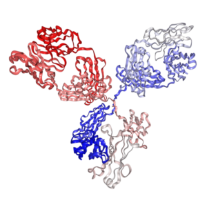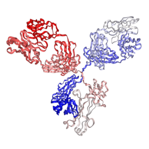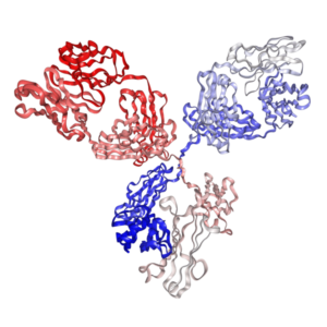
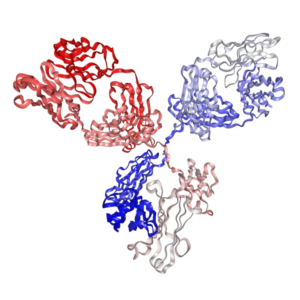

Anti-mouse CD4 Antibody (GK1.5) for Flow Cytometry | PA007494.h1Fs


$40.00 – $70.00
Recombinant rat IgG2b isotype controls and Recombinant human IgG1 isotype controls are available. Condition of sample preparation and optimal sample dilution should be determined experimentally by the investigator.
- Details & Specifications
- References
| Catalog No. | PA007494.h1Fs |
|---|---|
| Product Name | Recombinant Anti-mouse CD4 Monoclonal Antibody (Clone: GK1.5), Human IgG1 Fc Silent Kappa |
| Supplier Name | Syd Labs, Inc. |
| Brand Name | Syd Labs |
| Synonyms | L3T4, Ly-4, Leu-3, T4 |
| Clone | GK1.5. |
| Isotype | Human IgG1 kappa. |
| Source/Host | The anti-mouse CD4 monoclonal antibody (clone: GK1.5) for Flow Cytometry was produced in mammalian cells. |
| Specificity/Sensitivity | The recombinant GK1.5 antibody for Flow Cytometry specifically binds to mouse T cell receptor CD4. |
| Applications | Flow cytometry (FC), and Immunohistochemistry - Frozen (IHC-F). |
| Form Of Antibody | 0.2 uM filtered solution, pH 7.4, containing 0.09% sodium azide. |
| Conjugate | Non-conjugated. |
| Purity | >95% by SDS-PAGE under reducing conditions. |
| Shipping | The Recombinant Anti-mouse CD4 Monoclonal Antibody (Clone: GK1.5) for Flow Cytometry, Human IgG1 Fc Silent Kappa is shipped with ice pack. Upon receipt, store it immediately at the temperature recommended below. |
| Stability & Storage | Use a manual defrost freezer and avoid repeated freeze-thaw cycles. 12 months from date of receipt, 2 to 8°C as supplied. Do not freeze it. |
| Note | Recombinant human IgG1 isotype controls are available. Condition of sample preparation and optimal sample dilution should be determined experimentally by the investigator. |
| Order Offline | Phone: 1-617-401-8149 Fax: 1-617-606-5022 Email: message@sydlabs.com Or leave a message with a formal purchase order (PO) Or credit card. |
Description
PA007494.h1Fs: Recombinant Anti-mouse CD4 Monoclonal Antibody (Clone: GK1.5) for Flow Cytometry for Flow Cytometry, Human IgG1 Fc Silent Kappa
Syd Labs offers a premium Recombinant Anti-mouse CD4 Monoclonal Antibody (Clone: GK1.5), specifically engineered for flow cytometry applications. This human IgG1 Fc silent kappa antibody is designed for high specificity and low background, making it an essential tool for researchers in immunology, particularly those focused on T cell biology and immune system dynamics. With its recombinant production and advanced Fc silent feature, this antibody ensures precise and reliable detection of mouse CD4 in complex samples.
The GK1.5 clone is a gold-standard antibody for detecting mouse CD4, widely utilized in immunology research for its proven reliability. Originally derived from rat IgG2b kappa, this recombinant version has been engineered to feature a human IgG1 Fc silent kappa format, enhancing its versatility and performance in flow cytometry while minimizing cross-reactivity.
Enhance your flow cytometry studies with the Recombinant Anti-mouse CD4 Monoclonal Antibody (Clone: GK1.5) from Syd Labs. Order now to experience unparalleled specificity and reliability in your research. For inquiries or bulk orders, contact our expert team.
References of Anti-Mouse CD4 Monoclonal Antibody (Clone: GK1.5)
1. ImmunoPET Imaging of Murine CD4+ T Cells Using Anti-CD4 Cys-Diabody: Effects of Protein Dose on T Cell Function and Imaging
Freise, A. C., et al.Mol Imaging Biol. 2017 Aug;19(4):599-609. PMID: 27966069
“GK1.5 cys-diabody (cDb), a previously developed anti-mouse CD4 antibody fragment, was tested at different doses to assess its effects on positron emission tomography (PET) imaging and CD4+ T cell viability, proliferation, CD4 expression, and function. In vivo administration of GK1.5 cDb at the high dose of 40 µg caused a transient decrease in CD4 expression in spleen, blood, lymph nodes, and thymus, which recovered within 3 days post-injection; this effect was reduced, although not abrogated, when 2 µg was administered. Concentrations of GK1.5 cDb in excess of 25 nM significantly inhibited CD4+ T cell proliferation and interferon-γ production in vitro. We previously developed an anti-CD4 antibody fragment, GK1.5 cys-diabody (cDb), for immunoPET imaging of murine CD4+ T cells and described its use in monitoring CD4+ T cell reconstitution after hematopoietic stem cell transplantation. To investigate effects of GK1.5 cDb on the expression of CD4 by CD4+ T cells, we injected 40 µg GK1.5 cDb into C57BL/6 mice and measured surface CD4 expression by T cells in blood, spleen, ILN, and thymus after one or three days.”
2. Migrant memory B cells secrete luminal antibody in the vagina
Oh, J.E., et al.Nature. 2019 Jul;571(7763):122-126. PMID: 31189952
“Cells were surface stained with anti-CD3ε (145–2C11), anti-CD4 antibodies (GK1.5 and RM4–5), and anti-CD44 (IM7), and then fixed and permeabilized using a Cytofix/Cytoperm™ Fixation/Permeabilization kit according to the manufacturer’s instructions. To deplete CD4 T cells, mice were treated with 300 µg intravenously on day −4, −1 and 10 µg anti-mouse CD4 Ab (GK1.5) intravaginally for four consecutive days before virus challenge. Stained cells were sorted by FACS Aria (BD Biosciences, Mountain View, CA). CD45 +CD3ε +CD4 +, CD45 +CD3ε +CD8α +, CD45 +CD3ε −CD19 + cells (vagina) and CD45 +CD19 +IgD −IgG +MHCII +GL7 −CD38 + cells (spleen) were collected for further analysis. Anti-CD45.2 (104), anti-CD45.1 (A20), anti-CD45 (30-F11), anti-CD3ε (145–2C11), anti-CD4 antibodies (GK1.5 and RM4–5), anti-CD8α (53–6.7), anti-CD19 (6D5 and 1D3), anti-CD45R/B220 (RA3–6B2), anti-CD138 (281–2), anti-CD38 (90), anti-GL7 (GL7), anti-MHC class II (I-A/I-E, M5/114.15.2), anti-CD11c (N418), anti-IgD (11–26c.2a), anti-CD44 (IM7), anti-PD-L2 (TY25), anti-CD80 (16–10A1), anti-CD69 (H1.2F3), anti-CD86 (GL1) were purchased from…”
3. Effect of a CD4-depleting antibody on the development of Cryptococcus neoformans-induced allergic bronchopulmonary mycosis in mice
Arora, S., et al.Infect Immun. 2006 Jul;74(7):4339-48. PMID: 16790808
“Mice were treated with 300 μg of anti-CD4 monoclonal antibody (MAb) GK1.5 intraperitoneally on the day prior to C. neoformans inoculation and boosted with 100 μg of MAb at days 7 and 14. The efficiency of T-cell depletion was assessed by flow cytometric analysis using anti-CD4 Ab RM 4-4, which binds to a region of CD4 distinct from GK1.5. Mice were rendered CD4 + T-cell deficient by injecting them intraperitoneally with the anti-CD4 MAb GK1.5. In comparison, anti-CD4 Ab-treated animals had much less inflammation, including fewer macrophages and eosinophils (Fig. 2B). Histological sections of lungs from control and anti-CD4 Ab-treated mice at week 3 postinfection were stained with PAS, which stains mucus pink.”
4. Protocol for the isolation of mouse senescence-associated CD4+ T cells using flow cytometry and functional assays
Fukushima, Y., et al.STAR Protoc. 2023 Sep 15;4(3):102472. PMID: 37515759
“CD4 MicroBeads do not interfere with anti-CD4 Ab (clone GK1.5) in the staining panels (Tables 1 and 2) and thus are compatible. Anti-CD4 (GK1.5), FITC (1:200)” is listed in the Key resources table under Antibodies. Anti-CD4 (GK1.5), APC-Cy7 (1:200)” is listed in the Key resources table under Antibodies. CD4 | GK1.5 | FITC | 1:200″ is listed in Table 1 under Ab staining panel A to isolate CD153+ SA-T cells. CD4 | GK1.5 | APC-Cy7 | 1:200″ is listed in Table 2 under Ab staining panel B to isolate “enriched” CD153+ SA-T cells.”
5. T4+ T helper cell function in vivo: differential requirement for induction of antiviral cytotoxic T-cell and antibody responses
Ahmed, R., et al.J Virol. 1988 Jun;62(6):2102-2106. PMID: 2966865
“Two monoclonal antibodies (GKI.5 and RL172.4) directed against the L3T4 (T4) molecule were used for depleting T helper cells from mice. The rat anti-mouse hybridoma MAb GK1.5 was obtained from the American Type Culture Collection, Rockville, Md. For in vivo treatment with GK1.5, mice were injected intraperitoneally (i.p.) with a total of 1.5 mg of GK1.5 antibody. Phycoerythrin-conjugated anti-mouse L3T4 MAb (clone GK1.5), biotin-conjugated anti-mouse Lyt2 MAb (clone 53-6), and streptavidin-phycoerythrin were purchased from Becton Dickinson, Mountain View, Calif. Two injections of GK1.5 (300 µg each on days -3 and -1) caused >90% depletion of L3T4+ cells.”
For more technical references or data sheets regarding the Recombinant Anti-Mouse CD4 Monoclonal Antibody (Clone: GK1.5) formats, please contact our scientific support team at message@sydlabs.com or explore options via Syd Labs.
We provide recombinant anti-human CD4 antibody(gk1.5) for Flow Cytometry:
Recombinant Anti-mouse CD4 Monoclonal Antibody (Clone: GK1.5), Rat IgG2b Kappa
Recombinant Anti-mouse CD4 Monoclonal Antibody (Clone: GK1.5), Human IgG1 Fc Silent Kappa
We also provide the following recombinant anti-human CD4 monoclonal antibodies for flow cytometry:
Recombinant Anti-human CD4 monoclonal antibody (Clone: OKT4) for flow cytometry
Recombinant Anti-human CD4 monoclonal antibody (Clone: OKT4A) for flow cytometry
Recombinant Anti-human CD4 monoclonal antibody (Clone: 13B8.2) for flow cytometry
Recombinant Anti-human CD4 monoclonal antibody (Clone: SK3 / Anti-LEU 3a) for flow cytometry
Anti-mouse CD4 Antibody (GK1.5) for Flow Cytometry from: Recombinant Anti-mouse CD4 Monoclonal Antibody (Clone: GK1.5), Human IgG1 Fc Silent Kappa: PA007494.h1Fs Syd Labs
You may also like…
-
 Select optionsQuick View
-
 Select optionsQuick View