Anti-mouse PD-L1 Antibody (B7-H1, 10F.9G2.1) | PA007164.r2b
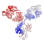

$150.00 – $900.00
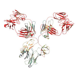
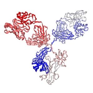
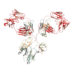
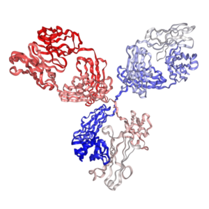
In Vivo Grade Recombinant Anti-mouse PD-L1 Rat IgG2b Kappa Monoclonal Antibody (Clone 10F.9G2.1). Recombinant anti-mouse PD L1 / B7-H1 monoclonal antibodies, which share the same variable region sequences with the rat anti-mouse PD L1 monoclonal antibody (clone number: 10F.9G2), are produced from mammalian cells and good for in vitro and in vivo studies.
- Details & Specifications
- References
| Catalog No. | PA007164.r2b |
|---|---|
| Product Name | In Vivo Grade Recombinant Anti-mouse PD-L1 Monoclonal Antibody (Clone 10F.9G2.1), Rat IgG2b Kappa |
| Supplier Name | Syd Labs, Inc. |
| Brand Name | Syd Labs |
| Synonyms | Programmed Death Ligand 1, B7-H1, CD274, 10F.9G2 antibody |
| Summary | The in vivo grade recombinant anti-mouse PD-L1 / B7-H1 monoclonal antibody (rat IgG2b kappa) was produced in mammalian cells. |
| Clone | 10F.9G2.1, the same variable region and constant region sequences as the rat anti-mouse PD-L1 monoclonal antibody (clone: 10F.9G2) |
| Isotype | Mouse IgG1, kappa |
| Applications | immunohistochemistry (IHC), Flow Cytometry (FC), and various in vitro and in vivo functional assays. |
| Immunogen | The original rat hybridoma (clone: 10F.9G2) was generated by immunizing rats with the mouse PD-L1 cDNA and mouse PD-L1 CHO transfectants. |
| Form Of Antibody | 0.2 μM filtered solution of 1x PBS. |
| Endotoxin | Less than 1 EU/mg of protein as determined by LAL method. |
| Purity | >95% by SDS-PAGE under reducing conditions. |
| Shipping | The in vivo grade recombinant anti-mouse PD L1/B7-H1 monoclonal antibodies are shipped with ice pack. Upon receipt, store it immediately at the temperature recommended below. |
| Stability & Storage | Use a manual defrost freezer and avoid repeated freeze-thaw cycles. 1 month from date of receipt, 2 to 8°C as supplied. 3 months from date of receipt, -20°C to -70°C as supplied. |
| Note | Recombinant anti-mouse PD L1/B7-H1 monoclonal antibodies, which share the same variable region sequences with the rat anti-mouse PD L1 monoclonal antibody (clone number: 10F.9G2), are produced from mammalian cells and good for in vitro and in vivo studies. |
| Order Offline | Phone: 1-617-401-8149 Fax: 1-617-606-5022 Email: message@sydlabs.com Or leave a message with a formal purchase order (PO) Or credit card. |
Description
PA007164.r2b: Recombinant Anti-mouse PD-L1 Antibody (Clone: 10F.9G2.1), Mouse IgG1, kappa, In Vivo Grade
The rat anti-mouse PD-L1 monoclonal antibody 10F.9G2 (rat IgG2b kappa) reacts with the mouse PD-L1 protein (programmed death ligand-1, B7-H1 or CD274 antibody), a member of the B7 family of the Ig superfamily. PD-1 has two ligands, PD-L1 and PD-L2. It has been shown that in mouse models of melanoma, tumor growth can be transiently arrested via treatment with the anti-mouse PD-1 and anti-mouse PD-L1 antibodies which block the interaction between the PD-L1 protein and its receptor PD-1 protein. The 10F.9G2 monoclonal antibody blocks the binding of the mouse PD-L1 protein to the mouse PD-1 protein.
Our recombinant 10F.9G2 antibodies have a part (variable regions) or complete amino acid sequences of the rat anti-mouse PD L1 monoclonal antibody (hybridoma clone name or number: 10F.9G2).
References of Recombinant Anti-Mouse PD-L1 Antibody (Clone: 10F.9G2)
1. The Antitumor Activity of Combinations of Cytotoxic Chemotherapy and Immune Checkpoint Inhibitors Is Model-Dependent.
Grasselly, et al.Front Immunol. 2018 Oct 9;9:2100. PMID: 30356816; PMCID: PMC6191484
“Immune checkpoint inhibitors (ICI), such as anti-PD-1 (Programmed cell death 1) or anti-PD-L1 (Programmed death-ligand 1) antibodies, are among the most important recent breakthroughs in oncology. …The PD-1/PD-L1 axis induces an inhibitory signal in T cells, and PD-1/PD-L1 pathway blockade restores T cell function resulting in increased proliferation and cytotoxic activity, subsequently improving anti-tumor immune response. …For this reason, it is crucial to develop new therapeutic approaches to enhance the therapeutic effects of PD-1/PD-L1 blockade, and to avoid resistance phenomena. …We performed an exploratory study of various combination regimens of chemotherapies with immune checkpoint blockers anti-PD-1 and anti-PD-L1, in several murine syngeneic preclinical models. …Remarkably the MVAC regimen had a different impact when combined with anti-PD-L1 Mab in the MB49 bladder model and in the MBT2 bladder model, supporting an influence of the model on the sensitivity to combination regimens.”
2. Highly pathological influenza A virus infection is associated with increased expression of PD-1 by functionally compromised virus-specific CD8+ T cells.
Rutigliano, et al.J Virol. 2014 Feb;88(3):1636-51. PMID: 24257598; PMCID: PMC3911675
“High-pathological infection was associated with increased PD-1 expression on influenza virus-specific CD8+ T cells, and blockade of PD-L1 in vivo led to reduced virus titers and increased CD8+ T cell numbers in high- but not low-pathological infection, though T cell functionality was not restored. …Blockade of PD-L1, one of two known ligands for PD-1, restores CD8+ T cell functionality and wholly improves the response to chronic lymphocytic choriomeningitis virus (LCMV) infection. …This expression was reversed by anti-PD-L1 treatment. Another study used a self-antigen system to show that PD-1 was expressed early on hemagglutinin-specific CD8+ T cells, and blockade of PD-L1 at the time of self-antigen encounter led to development of functional CD8+ T cells. …PD-L1 expression is also elevated on CD45+ MHCII? cells during infection with 104 EID50 of PR8. Interestingly, we found that at day 7, coexpression of PD-L1 and PD-L2 on MHCII+ CD45+ cells was spectacularly increased in x31-infected mice compared to PR8-infected mice. …Treatment with anti-PD-L1 antibody alone has been shown to rescue PD-1-induced exhaustion.”
3. Combining regulatory T cell depletion and inhibitory receptor blockade improves reactivation of exhausted virus-specific CD8+ T cells and efficiently reduces chronic retroviral loads.
Dietze, et al.PLoS Pathog. 2013;9(12):e1003798. PMID: 24339778; PMCID: PMC3868511
“The PD-1 receptor is a negative regulator of T cell proliferation and activation and is known to mediate suppressive functions when bound to its ligands PD-L1 or PD-L2. …Another group of animals was injected with monoclonal antibodies against PD-L1 and Tim-3 to block inhibitory receptor signaling on CD8+ T cells. …A 4-fold expansion of CD43+ CD8+ T cells was also found in mice treated with ?-PD-L1 and ?-TIM-3 but it was significantly lower than in the group of Treg depleted mice. …This suggested that ?-PD-L1 and ?-TIM-3 treatment might be more potent than ablation of Tregs in diminishing chronic virus loads. …However, this was only reflected in augmented FV-specific target cell killing when Treg depleted mice were compared to the animals receiving DT plus ?-PD-L1 and ?-TIM-3.”
4. The CTLA-4 and PD-1/PD-L1 inhibitory pathways independently regulate host resistance to Plasmodium-induced acute immune pathology.
Hafalla, et al.PLoS Pathog. 2012 Feb;8(2):e1002504. PMID: 22319445; PMCID: PMC3285150
“Accumulating data suggest that PD-1/PD-L1 signalling, and in some cases CTLA-4 signalling, is implicated in the T cell exhaustion that is seen in many chronic infections. …In vitro blockade of PD-1/PD-L1 pathway significantly increases CD8+ and CD4+ T cell function during HIV infection. …In vitro blockade of PD-1/PD-L1 and to a lesser extent PD-1/PD-L2 resulted in reversal of immune dysfunction in HCV. …Limited data are available for the PD-1/PD-L2 pathway during acute infections: PD-1/PD-L2 but not PD-1/PD-L1 blockade favours trypanosomatid growth in macrophages [35] and PD-L2 blockade enhances Th2 responses during Nippostrongylus infection [36]. …Thus, the CTLA-4 and PD-1/PD-L1 pathways seem to function very efficiently in BALB/c mice, maintaining the balance between immunity and immune pathology during the critical early stage of infection.”
5. PD-1/PD-L1 interactions inhibit antitumor immune responses in a murine acute myeloid leukemia model.
Zhang, et al.Blood. 2009 Aug 20;114(8):1545-52. PMID: 19417208; PMCID: PMC2729546
“In normal hosts, PD-1/PD-L1 interactions contribute to the maintenance of peripheral tolerance to self-antigens. …Conversely, PD-L1 mRNA is broadly expressed in tissues,7,8 and protein expression has been detected on many tumor cell types,15 and can be further induced by exposure to interferon (IFN)-?. Mounting evidence suggests that PD-L1 expression on solid tumor cells is capable of dampening antitumor T-cell responses. …Chen et al measured PD-L1 expression on bone marrow samples from patients with acute myeloid leukemia (AML) and found increasing levels upon disease progression, which was an independent negative prognostic factor for French-American-British type M5 AML. …To investigate if the PD-1/PD-L1 pathway promotes immune escape in a murine AML model, C57BL/6 or PD-1?/? mice were challenged intravenously (IV) with a highly lethal, syngeneic AML cell line, C1498, transduced to express green fluorescent protein (C1498.GFP) to allow monitoring of tumor burden. …These results confirm that the PD-1/PD-L1 pathway inhibits effective antitumor immune responses against murine AML, and support a rationale for clinical trials examining anti?PD-1 antibodies in patients with hematologic malignancies.”
For more technical references or data sheets regarding the Recombinant Anti-Mouse PD-L1 Monoclonal Antibody (Clone: 10F.9G2) formats, please contact our scientific support team at message@sydlabs.com.
Related Recombinant IgG Reference Antibodies:
Recombinant Mouse IgG1 Isotype Control Antibody and Mutants, In vivo Grade
Recombinant Mouse IgG2a Isotype Control Antibody and Mutants, In vivo Grade
Recombinant Mouse IgG2c Isotype Control Antibody and Mutants, In vivo Grade
Recombinant Rat IgG2a Isotype Control Antibody, In vivo Grade
Syd Labs provides the following anti-mouse PD-L1 / PD-1 antibodies:
Recombinant anti-mouse PD1 antibodies (Clone 29F.1A12.1), In vivo grade
Recombinant anti-mouse PD-1 antibodies (Clone RMP1-14.1), In vivo grade
Recombinant anti-mouse PD-L1 antibodies (Clone 10F.9G2.1), In vivo grade
Recombinant anti-mouse PD-1 / PD-1 bispecific antibodies (Clone RMP1-14.1 / 29F.1A12.1), In vivo grade
Recombinant anti-mouse PD-1 / PD-1 bispecific antibodies (Clone 29F.1A12.1 / RMP1-14.1), In vivo grade
Recombinant anti-mouse PD-1 / PD-L1 bispecific antibodies (Clone RMP1-14.1 / 10F.9G2.1), In vivo grade
Recombinant anti-mouse PD-L1 / PD-1 bispecific antibodies (Clone 10F.9G2.1 / RMP1-14.1), In vivo grade
Recombinant anti-mouse PD-1 / PD-L1 bispecific antibodies (Clone 29F.1A12.1 / 10F.9G2.1), In vivo grade
Recombinant anti-mouse PD-L1 / PD-1 bispecific antibodies (Clone 10F.9G2.1 / 29F.1A12.1), In vivo grade
Questions and Answers about anti-mouse PD-L1 antibody (Clone 10F.9G2):
Question: Do you produce Fc-silenced 10F.9G2 antibody?
Answer: Sure, we provide various recombinant Fc slient 10F.9G2 antibodies, such as mIgG2c LALAPG, mIgG2a LALAPG, and mIgG1 D265A. We also provide custom recombinant antibody production service to produce other engineered versions of recombinant 10F.9G2 antibodies.
Question: What is the difference among PA007164.r2b and PA007164.m2cLA?
Answer: PA007164.r2a is the recombinant anti-mouse PD L1 monoclonal antibody (rat IgG2b kappa, clone 10F.9G2.1) produced in CHO cells or HEK293 cells if needed. It has the same variable region and constant region sequences as the rat anti-mouse PD-L1 monoclonal antibody from the hybridoma clone of 10F.9G2. Rat antibodies may cause high immuogenicity in mice; thus, at least recombinant antibodies with mouse antibody constant regions should be used to replace the rat antibody constant regions. PA007164.m2cLA is the recombinant anti-mouse PD-L1 antibody (clone 10F.9G2.1) whose constant regions are mouse IgG2c LALAPG kappa.
Anti-mouse PD-L1 antibody (10F.9G2.1) from: Recombinant Anti-Myc-Tag Mouse IgG1 Monoclonal Antibody (clone 9E10): PA007164.r2b Syd Labs
You may also like…
-
 Select optionsQuick View
 - Select optionsQuick View
-
 Select optionsQuick View
 - Select optionsQuick View