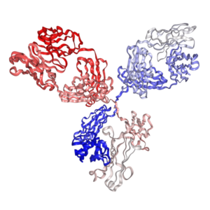

Anti-Mouse CD279 (PD-1) Antibody (Mouse IgG1, RMP1-14.1) | PA007162.m1


$400.00
In vivo anti-mouse CD279 (PD-1) antibody (mouse IgG1 κ, clone RMP1-14.1) — a recombinant mAb that pairs the identical antigen-binding variable region of the classic rat clone RMP1-14 with a wild-type mouse IgG1 kappa constant region.
As a low-effector isotype, it minimizes FcγR-mediated clearance of activated CD8⁺ T cells for functional PD-1 blockade without ADCC/CDC confounding.
Produced in mammalian cells; >95% purity, <1 EU/mg endotoxin.
- Details & Specifications
- References
- Other Formats
- Ushelf Advisor
| Catalog No. | PA007162.m1 |
|---|---|
| Product Name | In Vivo Grade Recombinant Anti-mouse PD-1 Monoclonal Antibody (Clone RMP1-14.1), Mouse IgG1 Kappa |
| Data Sheet | 📄 Download Product Data Sheet (PDF) |
| Supplier Name | Syd Labs, Inc. |
| Brand Name | Syd Labs |
| Synonyms | Anti-Mouse PD-1 antibody, Anti-CD279 antibody, Programmed cell death protein 1 antibody, Programmed death-1 antibody, Clone RMP1-14.1, Recombinant anti-mouse PD-1 mouse IgG1, In vivo anti-mouse PD-1 IgG1 |
| Summary | The In Vivo Grade Recombinant Anti-mouse PD-1 Mouse IgG1 Kappa Monoclonal Antibody (Clone RMP1-14.1) was produced in mammalian cells. |
| Clone | RMP1-14.1, the same variable region sequences as the rat anti-mouse PD-1 monoclonal antibody (clone number: RMP1-14) |
| Isotype | mouse IgG1, kappa |
| Applications | immunohistochemistry (IHC), Flow Cytometry (FC), and various in vitro and in vivo functional assays. |
| Immunogen | The original rat hybridoma (clone name: RMP1-14) was generated by immunizing rats with mouse PD-1-transfected BHK cells. |
| Form Of Antibody | 0.2 μM filtered solution of 1x PBS. |
| Endotoxin | Less than 1 EU/mg of protein as determined by LAL method. |
| Purity | >95% by SDS-PAGE under reducing conditions. |
| Shipping | The In Vivo Grade Recombinant Anti-mouse PD-1 Mouse IgG1 Kappa Monoclonal Antibody (Clone RMP1-14.1) are shipped with ice pack. Upon receipt, store it immediately at the temperature recommended below. |
| Stability & Storage | Use a manual defrost freezer and avoid repeated freeze-thaw cycles. 1 month from date of receipt, 2 to 8°C as supplied. 3 months from date of receipt, -20°C to -70°C as supplied. |
| Note | Recombinant mouse anti-mouse PD 1 / CD279 monoclonal antibodies, whose variable region sequences are murined from the rat anti-mouse PD-1 monoclonal antibody (clone number: RMP1-14), are produced from mammalian cells. The recombinant rat and chimeric mouse versions of the RMP1-14 antibody are also available. |
| Order Offline | Phone: 1-617-401-8149 Fax: 1-617-606-5022 Email: message@sydlabs.com Or leave a message with a formal purchase order (PO) Or credit card. |
Description
References of Anti-Mouse PD-1 Monoclonal Antibody (Clone: RMP1-14.1)
Selected publications on anti-mouse CD279 (PD-1) blockade and Fc effector function — including studies showing that FcγR engagement can deplete activated CD8⁺ T cells and blunt checkpoint blockade efficacy, the rationale for choosing a low-effector mouse IgG1 format such as PA007162.m1. Most studies used the parental RMP1-14 clone; where another isotype was used, the identical antigen-binding variable region makes the functional conclusions directly applicable to this format.
1. Macrophages Impair TLR9 Agonist Antitumor Activity through Interacting with the Anti-PD-1 Antibody Fc Domain
Simone Camelliti, et al. Cancers (Basel) 2021. doi: 10.3390/cancers13164081
“We evaluated the contribution of macrophages to the effect of combinatorial immunotherapeutic treatments based on TLR9 stimulation (with CpG-ODNs) and PD-1 blockade in an ovarian cancer preclinical model. We observed a strong reduction in the antitumor efficacy of a TLR9 agonist upon anti-PD-1 antibody administration. Specifically, we found that TLR9-stimulated macrophages, through interacting with the fragment crystallizable (Fc) domain of the anti-PD-1 antibody, acquire an immunoregulatory phenotype leading to dampening of CpG-ODN antitumor effect.”
2. Antibody-mediated depletion of programmed death 1-positive (PD-1+) cells
Yan Zhai, et al. J Control Release 2022. doi: 10.1016/j.jconrel.2022.07.010
“Anti-PD-1 antibodies are typically used to block inhibitory signals in dysfunctional T cells. Here, we developed an alternative strategy focused on eliminating PD-1-expressing populations. Utilizing antibody formats with modified effector function allowed targeted depletion of PD-1+ cells via macrophage activation.”
3. Decoupling FcRn and tumor contributions to elevated immune checkpoint inhibitor clearance in cancer cachexia
Thanh T. Vu, et al. Pharmacol Res 2024. doi: 10.1016/j.phrs.2023.107048
“Cancer cachexia accelerates the clearance of multiple monoclonal therapeutic antibodies, reducing exposure. We assessed the specific contributions of target burden and FcRn-mediated recycling dynamics. Our findings outline that cachexia altered antibody elimination tracks independently of typical target receptor distributions.”
4. Diphtheria toxin‐derived, anti‐PD‐1 immunotoxin, a potent and practical tool to selectively deplete PD‐1+ cells
Tao Zhang, et al. Protein Sci 2023. doi: 10.1002/pro.4741
“Investigating the roles of chronic PD-1+ populations requires reliable depletion methods. We engineered a diphtheria toxin-based immunotoxin architecture targeting mouse PD-1 structures. This molecule mediates rapid, highly specific systemic elimination of active PD-1-positive subgroups inside immunocompetent strains.”
For more technical references or data sheets regarding the Recombinant Anti-Mouse PD-1 Monoclonal Antibody (Clone: RMP1-14.1) formats, please contact our scientific support team at message@sydlabs.com.
Other Formats
All variants below share identical RMP1-14.1 variable region sequences and mouse PD-1 (CD279) binding specificity. They differ only in Fc isotype and Fc effector function — choose the format that matches your experimental readout and mouse strain.
| Catalog No. | Isotype | Fc Effector Function | Host / Reactivity | Applications |
|---|---|---|---|---|
| PA007162.r2a | Rat IgG2a, kappa | Intact (ADCC / CDC competent) | Rat / Mouse | IHC, FC, in vitro and in vivo functional assays |
| PA007162.m2a | Mouse IgG2a, kappa | Intact (ADCC / CDC competent) | Mouse / Mouse | IHC, FC, in vitro and in vivo functional assays |
| PA007162.m2c | Mouse IgG2c, kappa | Intact (ADCC / CDC competent) | Mouse / Mouse | IHC, FC, in vitro and in vivo functional assays |
| PA007162.m1 | Mouse IgG1, kappa | Low effector function | Mouse / Mouse | IHC, FC, in vitro and in vivo functional assays |
| PA007162.m2aLA | Mouse IgG2a-L234A L235A P329G (LALAPG), kappa | Fc-Silenced | Mouse / Mouse | IHC, FC, in vitro and in vivo functional assays, Fc-silenced neutralization |
| PA007162.m2cLA | Mouse IgG2c-L234A L235A P329G (LALAPG), kappa | Fc-Silenced | Mouse / Mouse | IHC, FC, in vitro and in vivo functional assays, Fc-silenced neutralization |
| PA007162.m1DA | Mouse IgG1-D265A, kappa | Fc-Silenced | Mouse / Mouse | IHC, FC, in vitro and in vivo functional assays, Fc-silent neutralization |
| PA007162.rt | Rabbit IgG | Rabbit Fc (non-mouse effector) | Rabbit / Mouse | IHC, FC, in vitro and in vivo functional assays |
How to choose a format: Use an intact-Fc format (r2a, m2a, m2c) when your readout requires full Fc-mediated effector activity (ADCC/CDC), and note that mouse IgG2c matches the C57BL/6 (Igh-1b) allotype commonly used in MC38 and B16-F10 models, while mouse IgG2a matches BALB/c (Igh-1a) backgrounds such as CT26. The low-effector wild-type mouse IgG1 format (m1) is suited to functional PD-1 blockade with minimal FcγR engagement, without full Fc silencing. Use an Fc-silenced format (m1DA, m2aLA, m2cLA) when you need pure PD-1 blockade without Fc-receptor engagement or ADCC/CDC confounding.
Ushelf Advisor
AI-generated answers, based on peer-reviewed literature and Ushelf product data. These FAQs cover isotype selection, Fc effector function comparison, experimental parameters, and handling for PA007162.m1 — our low-effector chimeric anti-mouse CD279 (PD-1) monoclonal antibody (mouse IgG1 κ, clone RMP1-14.1). This section is provided as a research reference. Please verify critical parameters — dosing, working dilutions, buffer conditions — against the primary references, or confirm with our technical support before finalizing an experimental design. Last updated: October 23, 2026.
What is the difference between PA007162.m1 and the rat IgG2a version (PA007162.r2a)?
PA007162.m1 is a recombinant chimeric anti-mouse CD279 (PD-1) antibody — it retains the identical rat-derived variable region of clone RMP1-14, paired with a wild-type mouse IgG1 kappa constant region instead of the rat IgG2a constant region of PA007162.r2a. The low-effector mouse IgG1 Fc is intended for functional PD-1 blockade without FcγR-mediated effector engagement.
Does PA007162.m1 mediate Fc effector function, and how does it differ from Fc-silenced formats?
PA007162.m1 is a wild-type mouse IgG1 isotype with low Fc effector function — it does not drive strong ADCC/CDC or FcγR-mediated depletion of activated CD8⁺ T cells. This distinguishes it from the Fc-silenced variants: PA007162.m1DA carries a D265A mutation (Fc-null), while PA007162.m2aLA and PA007162.m2cLA carry LALAPG mutations. Choose mouse IgG1 (m1) for a low-effector wild-type isotype, or m1DA when complete Fc silencing is required.
Is PA007162.m1 suitable for in vivo and functional applications?
Yes, this anti-mouse CD279 (PD-1) monoclonal antibody is produced in mammalian cells as an in vivo grade reagent with high purity (>95% by SDS-PAGE) and low endotoxin levels (<1 EU/mg by LAL method). It is validated for immunohistochemistry (IHC), flow cytometry (FC), and in vivo functional assays.
What specific clone characteristics and origin does PA007162.m1 have?
Designated as clone RMP1-14.1, it is derived from the original rat hybridoma (clone RMP1-14) generated by immunizing rats with mouse PD-1-transfected BHK cells, and shares identical variable region sequences with clone RMP1-14.
How do I choose between anti-mouse CD279 (PD-1) mouse IgG1 (PA007162.m1), IgG2a (PA007162.m2a) and IgG2c (PA007162.m2c)?
Choose by readout and host strain: mouse IgG1 (PA007162.m1) for low-effector functional PD-1 blockade; mouse IgG2a (PA007162.m2a) for BALB/c, C3H and DBA/2 (Igh-1a) backgrounds with intact Fc; mouse IgG2c (PA007162.m2c) for C57BL/6, C57BL/10 and NOD (Igh-1b) backgrounds. All formats share the identical RMP1-14.1 variable region and mouse PD-1 (CD279) binding specificity.
Which isotype control should I use with PA007162.m1?
A mouse IgG1, kappa isotype control matched to the same concentration and buffer composition is recommended for in vivo and flow cytometry controls. A matching isotype control is available for direct pairing with PA007162.m1.
What is the recommended in vivo dosage for PA007162.m1?
In mouse syngeneic tumor models, 200 µg per mouse administered intraperitoneally every 3 days is commonly used, with a frequently cited range of 200–500 µg per mouse. Optimal dosing and schedule should be determined for each specific animal model and study design.
How is PA007162.m1 formulated and supplied?
It is provided as a 0.2 µm filtered solution in 1× PBS, shipped with ice packs. Store in a manual defrost freezer at 2–8°C for short-term use (up to 1 month) or -20°C to -70°C for extended storage (up to 3 months), avoiding repeated freeze-thaw cycles.
You may also like…
-
 Select optionsQuick View
-
 Select optionsQuick View
-
 Select optionsQuick View
-
 Select optionsQuick View
-
 Select optionsQuick View