Anti-Mouse PD-1 (RMP1-14.1) In Vivo Antibody | PA007162.m2aLA
$150.00 – $700.00
In Vivo Recombinant Mouse Anti-Mouse PD 1 (RMP1-14.1) Antibody was produced in mammalian cells.
Applications: immunohistochemistry (IHC), Flow Cytometry (FC), and various in vitro and in vivo functional assays.
Endotoxin Level: Less than 1 EU/mg of protein as determined by LAL method.
- Details & Specifications
- References
- Ushelf Advisor
| Catalog No. | PA007162.m2aLA |
|---|---|
| Product Name | Anti-Mouse PD-1 (RMP1-14.1) In Vivo Antibody | PA007162.m2aLA |
| Supplier Name | Syd Labs, Inc. |
| Brand Name | Syd Labs |
| Synonyms | Mouse Anti-Mouse PD 1 Monoclonal Antibodies, Murinized Anti-Mouse PD 1 Monoclonal Antibodies |
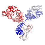
| Summary | The In Vivo Grade Recombinant Anti-mouse PD-1 Mouse IgG2a-L234A L235A P329G (LALAPG) Kappa Monoclonal Antibody (Clone RMP1-14.1) was produced in mammalian cells. |
| Clone | RMP1-14.1, the same variable region sequences as the rat anti-mouse PD-1 monoclonal antibody (clone number: RMP1-14) |
| Isotype | mouse IgG2a, kappa |
| Applications | immunohistochemistry (IHC), Flow Cytometry (FC), and various in vitro and in vivo functional assays. |
| Immunogen | The original rat hybridoma (clone name: RMP1-14) was generated by immunizing rats with mouse PD-1-transfected BHK cells. |
| Form Of Antibody | 0.2 μM filtered solution of 1x PBS. |
| Endotoxin | Less than 1 EU/mg of protein as determined by LAL method. |
| Purity | >95% by SDS-PAGE under reducing conditions. |
| Shipping | The In Vivo Grade Recombinant Anti-mouse PD-1 Mouse IgG2a-L234A L235A P329G (LALAPG) Kappa Monoclonal Antibody (Clone RMP1-14.1) are shipped with ice pack. Upon receipt, store it immediately at the temperature recommended below. |
| Stability & Storage | Use a manual defrost freezer and avoid repeated freeze-thaw cycles. 1 month from date of receipt, 2 to 8°C as supplied. 3 months from date of receipt, -20°C to -70°C as supplied. |
| Note | In Vivo Anti-mouse PD-1 (RMP1-14.1) Antibody, whose variable region sequences are murined from the rat anti-mouse PD-1 monoclonal antibody (clone number: RMP1-14), are produced from mammalian cells. The recombinant rat and chimeric mouse versions of the RMP1-14 antibody are also available. |
| Order Offline | Phone: 1-617-401-8149 Fax: 1-617-606-5022 Email: message@sydlabs.com Or leave a message with a formal purchase order (PO) Or credit card. |
Description
PA007162.m2aLA: Anti-Mouse PD-1 (RMP1-14.1) Antibody, Mouse IgG2a-L234A L235A P329G (LALAPG) Kappa,In Vivo Grade
The recombinant anti-mouse PD-1 (CD279) monoclonal antibody (Clone RMP1-14, Catalog No. PA007162.m2aLA) is a high-purity, in vivo-grade reagent engineered for precise blockade of the programmed death-1 (PD-1) immune checkpoint in mouse models. This non-depleting, Fc-silenced antibody features a mouse IgG2a isotype with L234A/L235A/P329G (LALAPG) mutations, eliminating Fcγ receptor binding and effector functions such as ADCC and CDC. As a result, it provides pure blocking-only activity without depleting PD-1-expressing cells (e.g., exhausted T cells), making it ideal for studies requiring clean PD-1/PD-L1/PD-L2 pathway inhibition.
Produced in mammalian cells for high specificity, consistency, and low endotoxin levels (<1 EU/mg, typical for in vivo-grade products), this antibody enhances anti-tumor immune responses by reversing T cell exhaustion and promoting effector functions in preclinical cancer immunotherapy research. It is widely applied in syngeneic tumor models (e.g., MC38, B16, CT26) on strains like C57BL/6, often in combination with anti-CTLA-4, radiotherapy, or chemotherapy.
References For Anti-Mouse PD-1 (RMP1-14.1) In Vivo Antibody:
1、Indoleamine 2,3-dioxygenase is a critical resistance mechanism in antitumor T cell immunotherapy targeting CTLA-4.
Holmgaard, R. B., et al. J Exp Med. 2013 Jul 1;210(7):1389-402. doi: 10.1084/jem.20130066. PMID: 23752227
This was also observed with antibodies targeting PD-1?PD-L1 and GITR. …Along these lines, we demonstrate that inhibition/absence of IDO in combination with therapies targeting immune checkpoints such as CTLA-4, PD-1/PD-L1, and GITR synergize to control tumor outgrowth and enhance overall survival in different tumor models. …In contrast to effector T cells, there was no significant increase in proliferation or up-regulation of PD-1 or ICOS by the tumor-infiltrating T reg cells with anti?CTLA-4 therapy. …Anti?PD-1/PD-L1 treatment delayed tumor progression in B16F10-bearing WT mice, but resulted in only modest improvement in survival. …However, anti?PD-1/PD-L1 treatment in IDO?/? mice resulted in significantly reduced tumor growth and improved survival.
Tags: bioactivity of anti-mouse PD-1 RMP1-14; anti-mouse PD-1 RMP1-14 of low endotoxin
2、Anti-PD-1 antibody therapy potently enhances the eradication of established tumors by gene-modified T cells.
John, L. B., et al. Clin Cancer Res. 2013 Oct 15;19(20):5636-46. doi: 10.1158/1078-0432.CCR-13-0458. PMID: 23873688
In this study, we show that blockade of the PD-1 immunosuppressive pathway using an anti-PD-1 antibody can significantly enhance the antitumor efficacy of genetically modified T cells expressing a chimeric antigen receptor (CAR). …The PD-1 pathway has emerged as another promising target for cancer therapy. …Engagement of the PD-1/PD-L1 pathway results in the phosphorylation of tyrosine-based motifs in the cytoplasmic tail of the PD-1 inhibitory receptor, which promotes the recruitment of SHP2 phosphatase leading to dephosphorylation of PI3K. …Recent trials using a fully human IgG4 PD-1 monocloncal antibody (mAb; BMS-936558) have reported durable clinical responses in patients with advanced melanoma, non?small cell lung, renal cell, as well as hematologic malignancies. …Hence, in this study, we hypothesized that CAR T-cell therapy in combination with PD-1 blockade may overcome PD-L1+ tumor immunosuppression, thereby leading to improved therapeutic efficacy.
Tags: anti-mouse PD-1 RMP1-14 mAb of low endotoxin; clone RMP1-14 of anti-mouse PD-1 antibody
Related Recombinant IgG Reference Antibodies:
Recombinant Mouse IgG1 Isotype Control Antibody and Mutants, In vivo Grade
Recombinant Mouse IgG2a Isotype Control Antibody and Mutants, In vivo Grade
Recombinant Mouse IgG2c Isotype Control Antibody and Mutants, In vivo Grade
Recombinant Rat IgG2a Isotype Control Antibody, In vivo Grade
Syd Labs provides the following anti-mouse PD-L1 / PD-1 antibodies:
Recombinant anti-mouse PD1 antibodies (Clone 29F.1A12.1), In vivo grade
Recombinant anti-mouse PD-1 antibodies (Clone RMP1-14.1), In vivo grade
Recombinant anti-mouse PD-L1 antibodies (Clone 10F.9G2.1), In vivo grade
Recombinant anti-mouse PD-1 / PD-1 bispecific antibodies (Clone RMP1-14.1 / 29F.1A12.1), In vivo grade
Recombinant anti-mouse PD-1 / PD-1 bispecific antibodies (Clone 29F.1A12.1 / RMP1-14.1), In vivo grade
Recombinant anti-mouse PD-1 / PD-L1 bispecific antibodies (Clone RMP1-14.1 / 10F.9G2.1), In vivo grade
Recombinant anti-mouse PD-L1 / PD-1 bispecific antibodies (Clone 10F.9G2.1 / RMP1-14.1), In vivo grade
Recombinant anti-mouse PD-1 / PD-L1 bispecific antibodies (Clone 29F.1A12.1 / 10F.9G2.1), In vivo grade
Recombinant anti-mouse PD-L1 / PD-1 bispecific antibodies (Clone 10F.9G2.1 / 29F.1A12.1), In vivo grade
Ushelf Advisor
Powered by AI: AI is experimental and still learning how to provide the best assistance. It may occasionally generate incorrect or incomplete responses. Please do not rely solely on its recommendations when making purchasing decisions or designing experiments.
Q: Is this anti-mouse PD-1 antibody low endotoxin and truly in vivo grade?
Yes, the recombinant anti-mouse PD-1/CD279 antibody (e.g., catalog variants like PA007162.m2aLA) qualifies as low endotoxin and in vivo grade based on standard scientific criteria used in preclinical immunology research.
Scientific Standards for In Vivo Use
Researchers require antibodies for in vivo PD-1 blockade in mouse models (e.g., syngeneic tumors or chronic infection) to have low endotoxin levels to prevent non-specific activation of innate immunity via TLR4 signaling, which could confound interpretation of adaptive immune responses (e.g., T cell exhaustion reversal).
Q: How effective is anti-mouse PD-1 (RMP1-14) in syngeneic tumor models like MC38 or B16?
The efficacy of anti-mouse PD-1 monoclonal antibody (clone RMP1-14) in syngeneic tumor models varies significantly depending on the tumor’s immunogenicity and microenvironment. RMP1-14 is one of the most widely used surrogates for human anti-PD-1 therapies (e.g., nivolumab) in preclinical studies, primarily functioning through blockade of the PD-1/PD-L1 axis to enhance T cell antitumor activity.
MC38 (Colorectal Carcinoma Model)
- MC38 is considered a highly immunogenic (“hot”) tumor model and is generally responsive to PD-1 blockade monotherapy with RMP1-14.
- Studies consistently demonstrate significant antitumor effects, including:
- Marked reduction in tumor growth (e.g., 2-3 fold decrease in relative tumor volume compared to controls).
- Delayed progression.
- Improved survival.
- Partial or complete tumor regressions in a substantial proportion of mice.
- MC38 is often classified as a benchmark responsive model for testing anti-PD-1 efficacy, with monotherapy achieving robust immune activation and serving as a positive control in many experiments.
B16 (Melanoma Model, Often B16-F10 Variant)
- B16 is a poorly immunogenic (“cold”) tumor model with low baseline T cell infiltration, showing limited responsiveness to PD-1 monotherapy with RMP1-14.
- As monotherapy, it typically results in only modest or negligible tumor growth delay, with minimal impact on overall survival and rare complete responses.
- Significant efficacy in B16 usually requires combination therapies (e.g., with tankyrase inhibitors, rapamycin nanoparticles, botulinum toxin, or other checkpoints/immunomodulators), where anti-PD-1 enhances synergistic effects leading to better tumor control and survival.
Responsiveness aligns with broader patterns in syngeneic models: Highly immunogenic tumors (e.g., MC38, CT26) respond well to anti-PD-1 monotherapy, while less immunogenic ones (e.g., B16) are resistant and better suited for testing combinations. Factors influencing outcomes include dosing (typically 200-250 μg i.p. every 3-5 days), timing, mouse strain (e.g., C57BL/6), and potential Fc-mediated effects of the rat IgG2a isotype. Results can vary across studies due to experimental variables, but the patterns above are consistent in reviews and primary publications.


