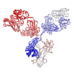
Anti-Mouse CD4 Antibody (GK1.5) | PA007200.r2b
$150.00 – $900.00
Recombinant rat IgG2b isotype controls are available. Condition of sample preparation and optimal sample dilution should be determined experimentally by the investigator.
- Details & Specifications
- References
| Catalog No. | PA007200.r2b |
|---|---|
| Product Name | Anti-Mouse CD4 Antibody (GK1.5) | PA007200.r2b |
| Supplier Name | Syd Labs, Inc. |
| Brand Name | Syd Labs |
| Synonyms | cluster of differentiation 4, CD4 |
| Summary | The anti-mouse CD4 monoclonal antibody (GK1.5) was produced in mammalian cells. |
| Uniprot ID | CD4 on UniProt.org |
| Gene ID | 12504 |
| Clone | GK1.5 |
| Isotype | Rat IgG2b kappa |
| Specificity/Sensitivity | The recombinant rat monoclonal antibody (clone: GK1.5), in vivo grade specifically binds to the mouse T cell receptor CD4. |
| Applications | ELISA, neutralization, functional assays such as bioanalytical PK and ADA assays, and those assays for studying biological pathways affected by the mouse CD4 protein. |
| Form Of Antibody | 0.2 uM filtered solution, pH 7.4, no stabilizers or preservatives. |
| Endotoxin | < 1 EU per 1 mg of the protein by the LAL method. |
| Purity | >95% by SDS-PAGE under reducing conditions and HPLC. |
| Shipping | The GK1.5 antibody is shipped with ice pack. Upon receipt, store it immediately at the temperature recommended below. |
| Stability & Storage | Use a manual defrost freezer and avoid repeated freeze-thaw cycles. 12 months from date of receipt, -20 to -70°C as supplied. 1 month from date of receipt, 2 to 8°C as supplied. |
| Note | Recombinant rat IgG2b isotype controls are available. Condition of sample preparation and optimal sample dilution should be determined experimentally by the investigator. |
| Order Offline | Phone: 1-617-401-8149 Fax: 1-617-606-5022 Email: message@sydlabs.com Or leave a message with a formal purchase order (PO) Or credit card. |
Description
PA007200.r2b: Recombinant Anti-mouse CD4 Monoclonal Antibody (Clone: GK1.5), Rat IgG2b Kappa, In vivo Grade
The anti mouse CD4 antibody (Clone GK1.5) specifically recognizes the 55 kDa CD4 glycoprotein, a crucial cell surface marker belonging to the immunoglobulin superfamily that serves as a coreceptor for MHC class II molecules. This rat IgG2b κ monoclonal antibody demonstrates high affinity for mouse CD4 expressed on thymocytes, helper T cells (including Th1, Th2, Th17 subsets), regulatory T cells (Treg), NK-T cells, and weakly on dendritic cells and macrophages. As a well-characterized CD4 antibody, GK1.5 plays essential roles in T cell development and function by stabilizing TCR-MHC II interactions through binding to conserved epitopes on the CD4 molecule.
This anti CD4 monoclonal antibody is particularly valuable for flow cytometry (recommended 0.5-1.0 µg per million cells), immunohistochemistry, and functional studies involving T cell activation or depletion in autoimmune disease models like experimental autoimmune encephalomyelitis (EAE). The GK1.5 clone competes with YTS 177 and YTS 191 antibodies for CD4 binding, making it an excellent tool for studying CD4-mediated signaling pathways, immune checkpoint regulation, and viral entry mechanisms (including HIV-1 and HHV-7). Available in multiple conjugates (FITC, PE, APC) or unconjugated formats, this mouse CD4-specific antibody remains a gold standard reagent for immunology research, enabling precise identification of CD4+ T cell subsets and evaluation of therapeutic interventions targeting CD4+ T cell function.
Syd Labs offers anti-mouse CD4 antibodies in various isotypes including Rat IgG2b, Mouse IgG2b, and Mouse IgG2a. The in vivo grade recombinant anti-mouse CD4 rat IgG2b kappa monoclonal antibody (Clone: GK1.5) of Syd Labs was produced in mammalian cells.
References for Recombinant Anti-mouse CD4 Antibodies (GK1.5):
1、Macrophage Migration Inhibitory Factor protects cancer cells from immunogenic cell death and impairs anti-tumor immune responses.
Balogh, K. N., et al. PLoS One. 2018 Jun 4;13(6):e0197702. doi: 10.1371/journal.pone.0197702. PMID: 29864117
“Beginning two days before 4T1 cell tumor implantation, mice were treated with intraperitoneal injection of an initial dose of 200ug/mouse of anti-CD4 (clone GK1.5, BioXCell) and anti-CD8 (clone 2.43, BioXCell) antibodies in PBS, followed by similar dosing with 100ug/mouse every 4 days throughout the course of tumor growth. …The Macrophage Migration Inhibitory Factor (MIF) is an inflammatory cytokine that is overexpressed in a number of cancer types, with increased MIF expression often correlating with tumor aggressiveness and poor patient outcomes. …We subsequently discovered that loss of MIF expression in 4T1 cells led to decreased cell numbers and increased apoptosis in vitro under reduced serum culture conditions. …Furthermore, we found that MIF depletion from the tumor cells resulted in greater numbers of activated intra-tumoral dendritic cells (DCs). …The Macrophage Migration Inhibitory Factor (MIF) was first described in the 1960’s as a T cell secreted factor capable of inhibiting the random migration of macrophages in vitro.”
2、IL-22 deficiency increases CD4 T cell responses to mucosal immunization.
Budda, S. A., et al. Vaccine. 2018 Jun 14;36(25):3694-3700. doi: 10.1016/j.vaccine.2018.05.011. PMID: 29739717
“Compared to wild-type control mice, IL-22 deficient mice had increased antigen-specific CD4 T cell responses to intrarectal immunization using a protein and cholera toxin adjuvant vaccine. …Mucosal surfaces are the first line of defense against numerous bacterial, viral and parasitic pathogens. …Upon antigen re-encounter, memory and effector T cells are directed to the mucosal membranes through tissue-specific homing receptors. …The cytokine is produced by both activated innate and adaptive lymphocytes. …In epithelial cells, recognition of IL-22 induces STAT3 activation, leading to pro-proliferative and anti-apoptotic pathways, which aids in epithelial barrier maintenance.”
3、Eradication of large established tumors in mice by combination immunotherapy that engages innate and adaptive immune responses.
Moynihan, K. D., et al. Nat Med. 2016 Dec;22(12):1402-1410. doi: 10.1038/nm.4200. PMID: 27775706
“Checkpoint blockade with antibodies specific for cytotoxic T lymphocyte–associated protein (CTLA)-4 or programmed cell death 1 (PDCD1; also known as PD-1) elicits durable tumor regression in metastatic cancer, but these dramatic responses are confined to a minority of patients. …Here we describe a combination immunotherapy that recruits a variety of innate and adaptive immune cells to eliminate large tumor burdens in syngeneic tumor models and a genetically engineered mouse model of melanoma; to our knowledge tumors of this size have not previously been curable by treatments relying on endogenous immunity. …Maximal antitumor efficacy required four components: a tumor-antigen-targeting antibody, a recombinant interleukin-2 with an extended half-life, anti-PD-1 and a powerful T cell vaccine. …Depletion experiments revealed that CD8+ T cells, cross-presenting dendritic cells and several other innate immune cell subsets were required for tumor regression. …Effective treatment induced infiltration of immune cells and production of inflammatory cytokines in the tumor, enhanced antibody-mediated tumor antigen uptake and promoted antigen spreading. …These results demonstrate the capacity of an elicited endogenous immune response to destroy large, established tumors and elucidate essential characteristics of combination immunotherapies that are capable of curing a majority of tumors in experimental settings typically viewed as intractable.”
4、TGFβ Is a Master Regulator of Radiation Therapy-Induced Antitumor Immunity.
Vanpouille-Box, C., et al. Cancer Res. 2015 Jun 1;75(11):2232-42. doi: 10.1158/0008-5472.CAN-14-3511. PMID: 25858148
“Briefly, TDLN cells were stained with anti-mouse CD4-PE and anti-mouse CD8-FITC (eBioscience), fixed, permeabilized (Foxp3 Fixation/Permeabilization Concentrate and Diluent kit, eBioscience), and stained with goat anti-phospho-Smad2/3 (Ser 423/425) followed by APC-labeled donkey anti-goat IgG (Santa Cruz Biotechnology). …Sections were incubated with 0.1% Tween 20 and 0.01% Triton X-100 for 20 minutes, followed by blocking with 4% rat serum in 4% BSA/PBS and staining with PE-Texas Red–rat anti-mouse CD4 or PE-rat anti-mouse CD8α (Caltag), and 5 μg/mL 4′,6-diamidino-2-phenylindole (Sigma). …Eliciting T-cell responses to a patient’s individual tumor remains a major challenge. …We hypothesized that TGFβ activity is a major obstacle hindering the ability of radiation to generate an in situ tumor vaccine. …Gene signatures associated with IFNγ and immune-mediated rejection were detected in tumors treated with radiation therapy and TGFβ blockade in combination but not as single agents.”
5、PD-1 Co-inhibitory and OX40 Co-stimulatory Crosstalk Regulates Helper T Cell Differentiation and Anti-Plasmodium Humoral Immunity.
Zander, R. A., et al. Cell Host Microbe. 2015 May 13;17(5):628-41. doi: 10.1016/j.chom.2015.03.007. PMID: 25891357
“Giemsa staining of thin blood smears was done in parallel. At the indicated times, mice were injected i.p. with 200 μg α-CD4 (GK1.5), 500 μg of α-IFN-γ (XMG1.2), 200 μg α-PD-L1 (10F.9G2), 50 μg of α-OX40 Ab (OX86), 200 μg α-PD-L1 and 50 μg α-OX40, or 200 μg α-PD-1 (RMP1-14) and 50 μg α-OX40, or equivalent amounts of rat IgG. …However, these beneficial effects of OX40 signaling are abrogated following coordinate blockade of PD-1 co-inhibitory pathways, which are also upregulated during malaria and associated with elevated parasitemia. …Our results show that targeting OX40 can enhance Plasmodium control and that crosstalk between co-inhibitory and co-stimulatory pathways in pathogen-specific CD4 T cells can impact pathogen clearance. …The differentiation and protective capacity of Plasmodium-specific T cells are regulated by both positive and negative signals during malaria, but the molecular and cellular details remain poorly defined. …To formally test whether CD4 T cells are necessary for the in vivo protective effects of α-OX40 during experimental malaria, we repeated our studies in CD4 T cell-depleted mice.”
6、Memory programming in CD8(+) T-cell differentiation is intrinsic and is not determined by CD4 help.
Kim, J., et al. Nat Commun. 2015 Aug 14;6:7994. doi: 10.1038/ncomms8994. PMID: 26272364
“Mice were injected i.p. with ascites fluid of anti-mouse CD4 monoclonal antibody (GK1.5) at 1 and 3 days before immunization. …Magnetic bead-based enrichment of T cells was performed as described previously. …Splenocytes from primed B6 mice (without J15 transfer) were stained with primary phycoerythrin (PE)-conjugated H60 tetramer and anti-PE magnetic microbeads to track the polyclonal H60-tetramer-binding CD8+ T cells. …In this study, we showed that impaired memory generation by CD8+ T cells activated in the absence of CD4 help is due to a failure to generate sufficient numbers of effector CD8 T cells at initial burst expansion on priming and the subsequent failure of efficient antigen clearance, and also that these T cells become eventually exhausted due to antigen persistence in our model. …Helper-deficient CD8+ T cells show reduced burst expansion, rapid peripheral egress, delayed antigen clearance and continuous activation, and are eventually exhausted.”
7、Innate immunological function of TH2 cells in vivo.
Guo, L., et al. Nat Immunol. 2015 Oct;16(10):1051-9. doi: 10.1038/ni.3244. PMID: 26322482
“Type 2 helper T cells (TH2 cells) produce interleukin 13 (IL-13) when stimulated by papain or house dust mite extract (HDM) and induce eosinophilic inflammation. …This innate response is dependent on IL-33 but not T cell antigen receptors (TCRs). …While type 2 innate lymphoid cells (ILC2 cells) are the dominant innate producers of IL-13 in naive mice, we found here that helminth-infected mice had more TH2 cells compared to uninfected mice, and thes e cells became major mediators of innate type 2 responses. …TH2 cells made important contributions to HDM-induced antigen-nonspecific eosinophilic inflammation and protected mice recovering from infection with Ascaris suum against subsequent infection with the phylogenetically distant nematode Nippostrongylus brasiliensis. …Our findings reveal a previously unappreciated role for effector TH2 cells during TCR-independent innate-like immune responses.”
8、IL-27 Signaling Is Crucial for Survival of Mice Infected with African Trypanosomes via Preventing Lethal Effects of CD4+ T Cells and IFN-γ.
Liu, G., et al. PLoS Pathog. 2015 Jul 29;11(7):e1005065. doi: 10.1371/journal.ppat.1005065. PMID: 26222157
“congolense were treated with depleting anti-mouse CD4 mAb, anti-mouse CD8 mAb, or rat IgG as control; and the course of infection, immune responses, and severity of liver damage were assessed. …Purified rat anti-mouse IL-10 receptor (IL-10R) mAb (Clone 1B1.3a), purified rat anti-mouse CD4 mAb (Clone GK1.5), purified rat anti-mouse CD8 (Clone 53–6.72), and purified rat anti-mouse IFN-γ mAb (Clone XMG1.2) were purchased from BioXCell (West Lebanon, NH). …African trypanosomes are extracellular protozoan parasites causing a chronic debilitating disease associated with a persistent inflammatory response. …Maintaining the balance of the inflammatory response via downregulation of activation of M1-type myeloid cells was previously shown to be crucial to allow prolonged survival. …Thus, our data identify IL-27 signaling as a novel pathway to prevent early mortality via inhibiting hyperactivation of CD4+ Th1 cells and their excessive secretion of IFN-γ during infection with African trypanosomes.”
9、Depletion of regulatory T cells in a hapten-induced inflammation model results in prolonged and increased inflammation driven by T cells.
Christensen, A. D., et al. Clin Exp Immunol. 2015 Mar;179(3):485-99. doi: 10.1111/cei.12466. PMID: 25302741
“Regulatory T cells (Tregs) are known to play an immunosuppressive role in the response of contact hypersensitivity (CHS), but neither the dynamics of Tregs during the CHS response nor the exaggerated inflammatory response after depletion of Tregs has been characterized in detail. …In this study we show that the number of Tregs in the challenged tissue peak at the same time as the ear-swelling reaches its maximum on day 1 after challenge, whereas the number of Tregs in the draining lymph nodes peaks at day 2. …As expected, depletion of Tregs by injection of a monoclonal antibody to CD25 prior to sensitization led to a prolonged and sustained inflammatory response which was dependent upon CD8 T cells, and co-stimulatory blockade with cytotoxic T lymphocyte antigen-4-immunoglobulin (CTLA-4-Ig) suppressed the exaggerated inflammation. …In contrast, blockade of the interleukin (IL)-10-receptor (IL-10R) did not further increase the exaggerated inflammatory response in the Treg-depleted mice. …Furthermore, depletion of Tregs enhanced the release of cytokines and chemokines locally in the inflamed ear and augmented serum levels of the systemic inflammatory mediators serum amyloid (SAP) and haptoglobin early in the response.”
10、Antibody Blockade of Semaphorin 4D Promotes Immune Infiltration into Tumor and Enhances Response to Other Immunomodulatory Therapies.
Evans, E. E., et al. Cancer Immunol Res. 2015 Jun;3(6):689-701. doi: 10.1158/2326-6066.CIR-14-0171. PMID: 25614511
“Other antibody clones, including anti–PD-1/MAb RMP1-14, anti-CD8/MAb 2.43, and anti-CD4/MAb GK1.5, were purchased from BioXCell and were verified to have <2 EU/mg endotoxin levels, ≥95% purity, and ≤5% high-molecular-weight species before in vivo use. …Cohorts of tumor-bearing mice were sacrificed at a predetermined endpoint, based on mean tumor volume (MTV) of the control group. …Semaphorin 4D (SEMA4D, CD100) and its receptor plexin-B1 (PLXNB1) are broadly expressed in murine and human tumors, and their expression has been shown to correlate with invasive disease in several human tumors. …In the setting of cancer, SEMA4D–PLXNB1 interactions have been reported to affect vascular stabilization and transactivation of ERBB2, but effects on immune-cell trafficking in the tumor microenvironment (TME) have not been investigated. …This orchestrated change in the tumor architecture was associated with durable tumor rejection in murine Colon26 and ERBB2+ mammary carcinoma models.”
We also provide other related Anti-Mouse CD4 antibody:
Anti-CD4 Antibody (mouse), clone GK1.5 | PA007200.m2c
Anti-mouse CD4 Antibody (Clone: GK1.5) | PA007200.m2b
Anti-mouse CD4 Antibody – GK1.5 | PA007200.m2a
Anti-mouse CD4 Monoclonal Antibody for Flow Cytometry (Clone: GK1.5) | PA007494.h1Fs
Anti-mouse CD4 Monoclonal Antibody for Flow Cytometry (Clone: GK1.5) | PA007494.r2b
We provide the following recombinant anti-human CD4 monoclonal antibodies:
Clenoliximab biosimilar, research grade, anti-human CD4 monoclonal antibody
Ibalizumab biosimilar, research grade, anti-human CD4 monoclonal antibody
Recombinant Anti-human CD4 monoclonal antibody (Clone: OKT4)
Recombinant Anti-human CD4 monoclonal antibody (Clone: OKT4A)
Recombinant Anti-human CD4 monoclonal antibody (Clone: 13B8.2)
Recombinant Anti-human CD4 monoclonal antibody (Clone: SK3 / Anti-LEU 3a)
We provide the following recombinant anti-mouse CD4 monoclonal antibodies:
Recombinant Anti-mouse CD4 monoclonal antibody (Clone: GK1.5)
We provide the following recombinant anti-human CD4 monoclonal antibodies for flow cytometry:
Recombinant Anti-human CD4 monoclonal antibody (Clone: OKT4) for flow cytometry
Recombinant Anti-human CD4 monoclonal antibody (Clone: OKT4A) for flow cytometry
Recombinant Anti-human CD4 monoclonal antibody (Clone: 13B8.2) for flow cytometry
Recombinant Anti-human CD4 monoclonal antibody (Clone: SK3 / Anti-LEU 3a) for flow cytometry
Anti-mouse CD4 Antibody (GK1.5) from: In vivo Grade Recombinant Anti-mouse CD4 Rat IgG2b Kappa Monoclonal Antibody (Clone: GK1.5): PA007200.r2b Syd Labs


