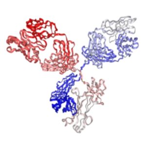
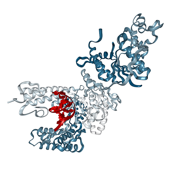
Biotinylated Anti-mouse PD1 Antibody (29F.1A12.1, Human IgG1 Fc Silent Kappa)
$80.00 – $160.00
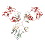
Recombinant mouse anti-mouse PD-1 / CD279 monoclonal antibody (Clone 29F.1A12, Biotinylated), engineered with a Human IgG1 Fc-Silent domain to minimize non-specific Fc receptor binding during flow cytometry and immune checkpoint analysis. Variable regions are derived from the classic rat anti-mouse PD-1 clone 29F.1A12 and recombinantly expressed in mammalian cells. For human T cell research, Syd Labs OKT3 antibodies are also available.
- Form & Conjugation: Biotinylated, Flow Cytometry Grade
- Applications: Flow Cytometry (FC), Immune Checkpoint Analysis, Functional Assays
- Alternative Formats: Non-biotinylated FC grade, unconjugated in vivo grade, and Syd Labs OKT3 antibodies are also available.
- Details & Specifications
- References
| Catalog No. | PA007734.h1Fs-b |
|---|---|
| Product Name | Biotinylated Recombinant Anti-mouse PD1 Monoclonal Antibody (Clone: 29F.1A12.1), Human IgG1 Fc Silent Kappa |
| Supplier Name | Syd Labs, Inc. |
| Brand Name | Syd Labs |
| Clone | 29F.1A12.1 |
| Isotype | Human IgG1 Fc Silent Kappa |
| Source/Host | The site-directed biotinylated anti-mouse PD1 monoclonal antibody (clone: 29F.1A12.1) was produced in mammalian cells. |
| Specificity/Sensitivity | The site-directed biotinylated recombinant 29F.1A12.1 antibody specifically binds to mouse PD1 protein. |
| Applications | Flow cytometry (FC) with various dye conjugation through streptavidin; conjugation to streptavidin agarose beads or streptavidin magnetic beads. |
| Form Of Antibody | 0.2 uM filtered solution, pH 7.4, no stabilizers or preservatives. |
| Conjugate | Biotin-conjugated |
| Purity | >95% by SDS-PAGE under reducing conditions and HPLC |
| Shipping | The 29F.1A12.1 antibody is shipped with ice pack. Upon receipt, store it immediately at the temperature recommended below. |
| Stability & Storage | Use a manual defrost freezer and avoid repeated freeze-thaw cycles. 12 months from date of receipt, -20 to -70°C as supplied. 1 month from date of receipt, 2 to 8°C as supplied. |
| Note | Recombinant rat IgG2b isotype controls and Recombinant human IgG1 isotype controls are available. Condition of sample preparation and optimal sample dilution should be determined experimentally by the investigator. |
| Order Offline | Phone: 1-617-401-8149 Fax: 1-617-606-5019 Email: message@sydlabs.com Or leave a message with a formal purchase order (PO) Or credit card. |
Description
PA007734.h1Fs-b: Biotinylated Recombinant Anti-mouse PD1 Monoclonal Antibody (Clone: 29F.1A12.1), Human IgG1 Fc Silent Kappa
Recombinant human IgG1 monoclonal antibody with site-directed biotinylation through the Avi tag at the C-terminus of the antibody heavy chain.
The rat anti-mouse PD-1 monoclonal antibody 29F.1A12 (rat IgG2a kappa) reacts with the mouse PD-1 protein (programmed death-1 or CD279) encoded by the mouse Pdcd1 gene, a member of the CD28 family of the Ig superfamily. PD-1 has two ligands, PD-L1 and PD-L2, both of which belong to the B7 family. It has been shown that in mouse models of melanoma, tumor growth can be transiently arrested via treatment with the anti-mouse PD-1 and anti-mouse PD-L1 antibodies which block the interaction between the PD-L1 protein and its receptor PD-1 protein. The 29F.1A12 monoclonal antibody blocks the binding of both the mouse PD-L1 protein and the mouse PD-L2 protein to the mouse PD-1 protein.
Our recombinant 29F.1A12 antibodies have a part (variable regions) or complete amino acid sequences of the rat anti-mouse PD-1 monoclonal antibody (hybridoma clone name or number: 29F.1A12).
References of Anti-Mouse PD-1 Monoclonal Antibody (Clone: 29F.1A12):
1. Anti-PD-1 therapy triggers Tfh cell-dependent IL-4 release to boost CD8 T cell responses in tumor-draining lymph nodes
Ruggiu, M., et al. J Exp Med. 2024;221(4):e20232104. PMID: 38417020
“Lymph node cells were stained ex vivo with a different anti-PD-1 mAb clone that did not interfere with the injected mAb to link in vivo labeling with PD-1 surface expression levels. …Representative FACS dot plot showing in vivo labeling by AF594-labeled anti-PD-1 mAb (clone RMP1-14) and ex vivo staining by anti-PD-1 antibody (clone 29F.1A12). …anti-mouse PD1-BX036 (purified clone 29F.1A12).”
Martins, C., et al. Sci Rep. 2022;12(1):12491. PMID: 35864188
“Similarly, in preclinical cancer models, several PD-1 blocking ab clones, including 29F.1A12 (anti-mouse PD-1 antibody), thwart tumor progression. …For example, TME-infiltrating macrophages and natural killer (NK) cells both express PD-1, the blockade of which by 29F.1A12 (anti-mouse PD-1 mAb) contributes to tumor growth suppression and prolonged survival. …29F.1A12 antibody recognizes PD-1 on B16-F10 and other murine tumor lines, suppresses cancer cell-intrinsic PD-1 receptor signaling, and inhibits resultant tumor growth in three-dimensional (3D) culture and tumor-bearing mice.”
3. Dying cells expose a nuclear antigen cross-reacting with anti-PD-1 monoclonal antibodies
Metzger, P., et al. Sci Rep. 2018;8(1):9040. PMID: 29892077
“In addition, PD-1 staining (clone 29F1A12) uncovered a FSC-H low population predominantly positive for PD-1. …PD-1 (clone 29F.1A12) and fixable viability dye (FVD) staining was assessed by flow cytometry. …PD-1 staining (clone 29F.1A12) in these cells could be due to binding of an intracellular PD-1 pool that becomes accessible in cells with leaky membranes. …Again, PD-1 mAb staining (clone 29F.1A12) was found in both PD-1 wild-type and knock-out cells in the dead cell fraction, indicative of unspecific binding. …We assessed PD-1 expression by flow cytometry and compared the two mainly used antibody clones (29F.1A12 and RMP1-14), which were also included in the above-mentioned study.”
Koyama, S., et al. Nat Commun. 2016;7:10501. PMID: 26883990
“Fluorescent conjugated anti-PD-1 antibody is the same clone (29F.1A12) as the therapeutic antibody. …PD-1-blocking antibody (clone 29F.1A12), TIM-3-blocking antibody (clone RMT3-23) and their isotype controls (clone 2A3) were injected intraperitoneally into the mice for therapeutic treatment (three times a week, 200 μg for PD1 and 100 μg for TIM-3 per dose).”
Pauken, K. E., et al. Cell Rep. 2020;31(13):107827. PMID: 32610128
“WT B6 mice were treated with anti-PD-1 antibody (clone 29F.1A12) or isotype control antibody (rat IgG2a) on days -1, 2, and 5 and harvested on day 8 p.i., or treated on days -1, 2, 5, and 8 and harvested on day 60+ p.i. PD-L1/L2 DKO mice were included as a control. …For in vivo antibody blockade experiments, WT C57BL/6 mice were given 200 μg per mouse per injection of rat anti-PD-1 antibody (clone 29F.1A12) or isotype control antibody (Rat IgG2a, clone 2A3) on days -1, 2, and 5 p.i. and analyzed at day 8 p.i., or on days -1, 2, 5, and 8 p.i. and analyzed at day 60+ p.i.”
For more technical references or data sheets regarding the Recombinant Anti-Mouse PD-L1 Monoclonal Antibody (Clone: 10F.9G2) formats, please contact our scientific support team at message@sydlabs.com.
Syd Labs provides the following in vivo grade recombinant anti-mouse PD-L1 / PD-1 monoclonal antibodies:
In vivo grade recombinant anti-mouse PD1 monoclonal antibodies (Clone 29F.1A12)
In vivo grade recombinant anti-mouse PD-1 monoclonal antibodies (Clone RMP1-14)
In vivo grade recombinant anti-mouse PD-L1 monoclonal antibodies (Clone 10F.9G2)
Syd Labs provides the following in vivo grade recombinant anti-mouse PD-1 / PD-L1 bispecific antibodies:
In vivo grade recombinant anti-mouse PD-1 / PD-1 bispecific antibodies (Clone RMP1-14 / 29F.1A12)
In vivo grade recombinant anti-mouse PD-1 / PD-1 bispecific antibodies (Clone 29F.1A12 / RMP1-14)
In vivo grade recombinant anti-mouse PD-1 / PD-L1 bispecific antibodies (Clone RMP1-14 / 10F.9G2)
In vivo grade recombinant anti-mouse PD-L1 / PD-1 bispecific antibodies (Clone 10F.9G2 / RMP1-14)
In vivo grade recombinant anti-mouse PD-1 / PD-L1 bispecific antibodies (Clone 29F.1A12 / 10F.9G2)
In vivo grade recombinant anti-mouse PD-L1 / PD-1 bispecific antibodies (Clone 10F.9G2 / 29F.1A12)
Syd Labs provides the following recombinant anti-mouse PD-1 / PD-L1 monoclonal antibodies for flow cytometry:
Recombinant Anti-mouse PD-1 monoclonal antibodies (Clone: RMP1-14) for flow cytometry
Recombinant Anti-mouse PD-1 monoclonal antibodies (Clone: 29F.1A12) for flow cytometry
Recombinant Anti-mouse PD-L1 monoclonal antibodies (Clone: 10F.9G2) for flow cytometry
Recombinant PD1 and PDL1 Proteins:
Recombinant Human PD-1/PDCD1 Proteins
Biotinylated Recombinant Human PD-1/PDCD1 Proteins
Recombinant Mouse PD-1/PDCD1 Proteins
Biotinylated Recombinant Mouse PD-1/PDCD1 Proteins
Recombinant Cynomolgus PD-1/PDCD1 Proteins
Recombinant Canine PD-1/PDCD1 Proteins
Biotinylated Cynomolgus/Rhesus macaque PD-L1 Protein
Biotinylated Human PD-L1 Protein
Biotinylated Mouse PD-L1 Protein
Cynomolgus PD-L1 Protein
Cynomolgus/Rhesus macaque PD-L1 Protein
Human PD-L1/B7-H1 Protein
Mouse PD-L1/B7-H1 Protein
You may also like…
-
 Select optionsQuick View
-
 Select optionsQuick View
-
 Select optionsQuick View
-
 Select optionsQuick View