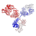
Anti Mouse CD3 (17A2) , In vivo | PA007199.m2c
$400.00
Recombinant rat IgG2b isotype controls are available. Condition of sample preparation and optimal sample dilution should be determined experimentally by the investigator.
- Details & Specifications
- References
| Catalog No. | PA007199.m2c |
|---|---|
| Product Name | Anti Mouse CD3 (17A2) , In vivo | PA007199.m2c |
| Supplier Name | Syd Labs, Inc. |
| Brand Name | Syd Labs |
| Clone | 17A2. |
| Isotype | Mouse IgG2c Kappa |
| Source/Host | The anti-mouse CD3 monoclonal antibody (clone: 17A2) was produced in mammalian cells. |
| Specificity/Sensitivity | The in vivo grade recombinant rat monoclonal antibody (clone: 17A2) specifically binds to the mouse T cell receptor CD3. |
| Applications | ELISA, neutralization, functional assays such as bioanalytical PK and ADA assays, and those assays for studying biological pathways affected by the mouse CD3 protein. |
| Form Of Antibody | 0.2 uM filtered solution, pH 7.4, no stabilizers or preservatives. |
| Endotoxin | < 1 EU per 1 mg of the protein by the LAL method. |
| Purity | >95% by SDS-PAGE under reducing conditions and HPLC. |
| Shipping | The In vivo Grade Recombinant Anti-mouse CD3 Monoclonal Antibody (Clone: 17A2), Mouse IgG2c Kappa is shipped with ice pack. Upon receipt, store it immediately at the temperature recommended below. |
| Stability & Storage | Use a manual defrost freezer and avoid repeated freeze-thaw cycles. 12 months from date of receipt, -20 to -70°C as supplied. 1 month from date of receipt, 2 to 8°C as supplied. |
| Note | Recombinant rat IgG2b isotype controls are available. Condition of sample preparation and optimal sample dilution should be determined experimentally by the investigator. |
| Order Offline | Phone: 1-617-401-8149 Fax: 1-617-606-5022 Email: message@sydlabs.com Or leave a message with a formal purchase order (PO) Or credit card. |
Description
PA007199.m2c: Recombinant Anti-Mouse CD3 Monoclonal Antibody(17A2) , Mouse IgG2c Kappa, In vivo Grade
References for Anti-Mouse CD3 (17A2) ,In vivo:
1、A TNIP1-driven systemic autoimmune disorder with elevated IgG4
Arti Medhavy,et al.Nat Immunol. 2024.PMCID: PMC11362012
“Whole-exome sequencing of two unrelated kindreds with Anti-Mouse CD3 systemic autoimmune disease featuring antinuclear antibodies with IgG4 elevation uncovered an identical ultrarare heterozygous TNIP1Q333P variant segregating with disease. Mice with the orthologous Q346P variant developed antinuclear autoantibodies, salivary gland inflammation, elevated IgG2c, spontaneous germinal centers and expansion of age-associated B cells, plasma cells and follicular and extrafollicular helper T cells. B cell phenotypes were cell-autonomous and rescued by ablation of Toll-like receptor 7 (TLR7) or MyD88. The variant increased interferon-β without altering nuclear factor kappa-light-chain-enhancer of activated B cells signaling, and impaired MyD88 and IRAK1 recruitment to autophagosomes. Additionally, the Q333P variant impaired TNIP1 localization to damaged mitochondria and mitophagosome formation. Damaged mitochondria were abundant in the salivary epithelial cells of Tnip1Q346P mice. These findings suggest that TNIP1-mediated autoimmunity may be a consequence of increased TLR7 signaling due to impaired recruitment of downstream signaling molecules and damaged mitochondria to autophagosomes and may thus respond to TLR7-targeted therapeutics.”
2、DDO-adjuvanted influenza A virus nucleoprotein mRNA vaccine induces robust humoral and cellular type 1 immune responses and protects mice from challenge
Victoria Gnazzo,et al.mBio. 2024.PMCID: PMC11796404
“A challenge in viral vaccine development is to produce vaccines that generate both neutralizing antibodies to prevent infection and cytotoxic CD8+ T-cells that target conserved viral proteins and can eliminate infected cells to control virus spread. mRNA technology offers an opportunity to design vaccines based on conserved CD8-targeting epitopes, but achieving robust antigen-specific CD8+ T-cells remains a challenge. Here, we tested the viral-derived oligonucleotide DDO268 as an adjuvant in the context of a model influenza A virus (IAV) nucleoprotein (NP) mRNA vaccine in C57BL/6 mice. DDO268 when co-packaged with mRNA in lipid nanoparticles is sensed by RIG I-like receptors and safely induces local type I interferon (IFN) production followed by dendritic cells type 1 activation and migration to the draining lymph nodes. This early response triggered by DDO268 improved the generation of IgG2c antibodies and antigen-specific Th1 CD4+ and CD8+ T-cells (IFNγ+TNFα+IL2+) that provided enhanced protection against lethal IAV challenge. In addition, the inclusion of DDO268 reduced the antigen dose required to achieve protection. These results highlight the potential of DDO268 as an effective mRNA vaccine adjuvant and show that an IAV NP mRNA/DDO268 vaccine is a promising approach for generating protective immunity against conserved internal IAV epitopes.”
3、Reduced SARS-CoV-2 mRNA vaccine immunogenicity and protection in mice with diet-induced obesity and insulin resistance
Timothy R O’Meara,et al.J Allergy Clin Immunol. 2024.PMCID: PMC10841117
“Background:
Obesity and Type 2 Diabetes Mellitus (T2DM) are associated with an increased risk of severe outcomes from infectious diseases, including COVID-19. These conditions are also associated with distinct responses to immunization, including an impaired response to widely used SARS-CoV-2 mRNA vaccines.
Objective:
To establish a connection between reduced immunization efficacy via modeling the effects of metabolic diseases on vaccine immunogenicity that is essential for the development of more effective vaccines for this distinct vulnerable population.
Methods:
We utilized a murine model of diet-induced obesity and insulin resistance to model the effects of comorbid T2DM and obesity on vaccine immunogenicity and protection.
Results:
Mice fed a high-fat diet (HFD) developed obesity, hyperinsulinemia, and glucose intolerance. Relative to mice fed a normal diet (ND), HFD mice vaccinated with a SARS-CoV-2 mRNA vaccine exhibited significantly lower anti-spike IgG titers, predominantly in the IgG2c subclass, associated with a lower type 1 response, along with a 3.83-fold decrease in neutralizing titers. Furthermore, enhanced vaccine-induced spike-specific CD8+ T cell activation and protection from lung infection against SARS-CoV-2 challenge were seen only in ND mice but not in HFD mice.
Conclusion:
We demonstrate impaired immunity following SARS-CoV-2 mRNA immunization in a murine model of comorbid T2DM and obesity, supporting the need for further research into the basis for impaired anti-SARS-CoV-2 immunity in T2DM and investigation of novel approaches to enhance vaccine immunogenicity among those with metabolic diseases.”
4、Alterations in germinal center formation and B cell activation during severe Orientia tsutsugamushi infection in mice
Casey Gonzales,et al.PLoS Negl Trop Dis. 2023.PMCID: PMC10191367
“Scrub typhus is a poorly studied but life-threatening disease caused by the intracellular bacterium Orientia tsutsugamushi (Ot). Cellular and humoral immunity in Ot-infected patients is not long-lasting, waning as early as one-year post-infection; however, its underlying mechanisms remain unclear. To date, no studies have examined germinal center (GC) or B cell responses in Ot-infected humans or experimental animals. This study was aimed at evaluating humoral immune responses at acute stages of severe Ot infection and possible mechanisms underlying B cell dysfunction. Following inoculation with Ot Karp, a clinically dominant strain known to cause lethal infection in C57BL/6 mice, we measured antigen-specific antibody titers, revealing IgG2c as the dominant isotype induced by infection. Splenic GC responses were evaluated by immunohistology, co-staining for B cells (B220), T cells (Anti-Mouse CD3), and GCs (GL-7). Organized GCs were evident at day 4 post-infection (D4), but they were nearly absent at D8, accompanied by scattered T cells throughout splenic tissues. Flow cytometry revealed comparable numbers of GC B cells and T follicular helper (Tfh) cells at D4 and D8, indicating that GC collapse was not due to excessive death of these cell subtypes at D8. B cell RNAseq analysis revealed significant differences in expression of genes associated with B cell adhesion and co-stimulation at D8 versus D4. The significant downregulation of S1PR2 (a GC-specific adhesion gene) was most evident at D8, correlating with disrupted GC formation. Signaling pathway analysis uncovered downregulation of 71% of B cell activation genes at D8, suggesting attenuation of B cell activation during severe infection. This is the first study showing the disruption of B/T cell microenvironment and dysregulation of B cell responses during Ot infection, which may help understand the transient immunity associated with scrub typhus.”
5、Bank1 modulates the differentiation and molecular profile of key B cell populations in autoimmunity
Gonzalo Gómez Hernández,et al.JCI Insight. 2024.PMCID: PMC11466193
“This study aimed at defining the role of the B cell adaptor protein BANK1 in the appearance of age-associated B cells (ABCs) in 2 SLE mouse models (TLR7.tg6 and imiquimod-induced mice), crossed with Bank1–/– mice. The absence of Bank1 led to a significant reduction in ABC levels, also affecting other B cell populations. To gain deeper insights into their differentiation pathway and the effect of Bank1 on B cell populations, a single-cell transcriptome assay was performed. In the TLR7.tg6 model, we identified 10 clusters within B cells, including an ABC-specific cluster that was decreased in Bank1-deficient mice. In its absence, ABCs exhibited an antiinflammatory gene expression profile, while being proinflammatory in Bank1-sufficient lupus-prone mice. Trajectory analyses revealed that ABCs originated from marginal zone and memory-like B cells, ultimately acquiring transcriptional characteristics associated with atypical memory cells and long-lived plasma cells. Also, Bank1 deficiency normalized the presence of naive B cells, which were nearly absent in lupus-prone mice. Interestingly, Bank1 deficiency significantly reduced a distinct cluster containing IFN-responsive genes. These findings underscore the critical role of Bank1 in ABC development, affecting early B cell stages toward ABC differentiation, and the presence of IFN-stimulated gene–containing B cells, both populations determinant for autoimmunity.”
6、Spatial proteogenomics reveals distinct and evolutionarily conserved hepatic macrophage niches
Martin Guilliams,et al.Cell. 2022.PMCID: PMC8809252
“The liver is the largest solid organ in the body, yet it remains incompletely characterized. Here we present a spatial proteogenomic atlas of the healthy and obese human and murine liver combining single-cell CITE-seq, single-nuclei sequencing, spatial transcriptomics, and spatial proteomics. By integrating these multi-omic datasets, we provide validated strategies to reliably discriminate and localize all hepatic cells, including a population of lipid-associated macrophages (LAMs) at the bile ducts. We then align this atlas across seven species, revealing the conserved program of bona fide Kupffer cells and LAMs. We also uncover the respective spatially resolved cellular niches of these macrophages and the microenvironmental circuits driving their unique transcriptomic identities. We demonstrate that LAMs are induced by local lipid exposure, leading to their induction in steatotic regions of the murine and human liver, while Kupffer cell development crucially depends on their cross-talk with hepatic stellate cells via the evolutionarily conserved ALK1-BMP9/10 axis.”
7、Inflammatory Type 2 cDCs Acquire Features of cDC1s and Macrophages to Orchestrate Immunity to Respiratory Virus Infection
Cedric Bosteels,et al.Immunity. 2020.PMCID: PMC7207120
“The phenotypic and Anti-Mouse CD3 functional dichotomy between IRF8+ type 1 and IRF4+ type 2 conventional dendritic cells (cDC1s and cDC2s, respectively) is well accepted; it is unknown how robust this dichotomy is under inflammatory conditions, when additionally monocyte-derived cells (MCs) become competent antigen-presenting cells (APCs). Using single-cell technologies in models of respiratory viral infection, we found that lung cDC2s acquired expression of the Fc receptor CD64 shared with MCs and of IRF8 shared with cDC1s. These inflammatory cDC2s (inf-cDC2s) were superior in inducing CD4+ T helper (Th) cell polarization while simultaneously presenting antigen to CD8+ T cells. When carefully separated from inf-cDC2s, MCs lacked APC function. Inf-cDC2s matured in response to cell-intrinsic Toll-like receptor and type 1 interferon receptor signaling, upregulated an IRF8-dependent maturation module, and acquired antigens via convalescent serum and Fc receptors. Because hybrid inf-cDC2s are easily confused with monocyte-derived cells, their existence could explain why APC functions have been attributed to MCs.”
8、A booster dose enhances immunogenicity of the COVID-19 vaccine candidate ChAdOx1 nCoV-19 in aged mice
Alyssa Silva-Cayetano,et al.Med. 2021.PMCID: PMC7833318
“Background
The spread of SARS-CoV-2 has caused a worldwide pandemic that has affected almost every aspect of human life. The development of an effective COVID-19 vaccine could limit the morbidity and mortality caused by infection and may enable the relaxation of social-distancing measures. Age is one of the most significant risk factors for poor health outcomes after SARS-CoV-2 infection; therefore, it is desirable that any new vaccine candidates elicit a robust immune response in older adults.
Methods
Here, we use in-depth immunophenotyping to characterize the innate and adaptive immune response induced upon intramuscular administration of the adenoviral vectored ChAdOx1 nCoV-19 (AZD-1222) COVID-19 vaccine candidate in mice.
Findings
A single vaccination generates spike-specific Th1 cells, Th1-like Foxp3+ regulatory T cells, polyfunctional spike-specific CD8+ T cells. and granzyme-B-producing CD8 effectors. Spike-specific IgG and IgM are generated from both the early extrafollicular antibody response and the T follicular helper cell-supported germinal center reaction, which is associated with the production of virus-neutralizing antibodies. A single dose of this vaccine generated a similar type of immune response in aged mice but of a reduced magnitude than in younger mice. We report that a second dose enhances the immune response to this vaccine in aged mice.
Conclusions
This study shows that ChAdOx1 nCoV-19 induces both cellular and humoral immunity in adult and aged mice and suggests a prime-boost strategy is a rational approach to enhance immunogenicity in older persons.
Funding
This study was supported by BBSRC, Lister institute of Preventative Medicine, EPSRC VaxHub, and Innovate UK.”
9、Increased number of T cells and exacerbated inflammatory pathophysiology in a human IgG4 knock-in MRL/lpr mouse model
Yoshie Gon,et al.PLoS One. 2023.PMCID: PMC9916631
“Immunoglobulin (Ig) G4 is an IgG subclass that can exhibit inhibitory functions under certain conditions because of its capacity to carry out Fab-arm exchange, inability to form immune complexes, and lack of antibody-dependent and complement-dependent cytotoxicity. Although several diseases have been associated with IgG4, its role in the disease pathogeneses remains unclear. Since mice do not express an IgG subclass that is identical to the human IgG4 (hIgG4), we generated hIGHG4 knock-in (KI) mice and analyzed their phenotypes. To preserve the rearrangement of the variable, diversity, and joining regions in the IGH gene, we transfected a constant region of the hIGHG4 gene into C57BL/6NCrSlc mice by using a gene targeting method. Although the mRNA expression of hIGHG4 was detected in the murine spleen, the serum level of the hIgG4 protein was low in C57BL/6-IgG4KI mice. To enhance the production of IgG4, we established an MRL/lpr-IgG4KI mice model by backcrossing. These mice showed a high IgG4 concentration in the sera and increased populations of IgG4-positive plasma cells and CD3+B220+CD138+ T cells in the spleen. Moreover, these mice showed aggravated inflammation in organs, such as the salivary glands and stomach. The MRL/lpr-IgG4KI mouse model established in the present study might be useful for studying IgG4-related disease, IgG4-type antibody-related diseases, and allergic diseases.”
10、Remarkably Robust Antiviral Immune Response Despite Combined Deficiency in Caspase-8 and RIPK3
Yanjun Feng,et al.J Immunol. 2019.PMCID: PMC6211196
“Caspase-8 (Casp8)-mediated signaling triggers extrinsic apoptosis while suppressing receptor interacting protein kinase (RIPK)3-dependent necroptosis. Even though Casp8 is dispensable for the development of innate and adaptive immune compartments in mice, the importance of this proapoptotic protease in the orchestration of immune response to pathogens remains to be fully explored. Here, Casp8−/−Ripk3−/− C57BL/6 mice show robust innate and adaptive immune responses to the natural mouse pathogen, murine cytomegalovirus. When young, these mice lack lpr-like lymphoid hyperplasia and accumulation of either B220+Anti-Mouse CD3+ or B220−CD3+ CD4+ and CD8+ T cells with increased numbers of immature myeloid cells that are evident in older mice. Dendritic cell activation and cytokine production drive both NK and T cell responses to control viral infection in these mice, suggesting that Casp8 is dispensable to the generation of antiviral host defense. Curiously, NK and T cell expansion is amplified, with greater numbers observed by 7 days post infection compared to either Casp8+/−Ripk3−/− or wild type (Casp8+/+Ripk3+/+) littermate controls. Casp8 and RIPK3 are natural targets of virus-encoded cell death suppressors that prevent infected cell apoptosis and necroptosis, respectively. It is clear from the current studies that the initiation of innate immunity and the execution of cytotoxic lymphocyte functions are all preserved despite the absence of Casp8 in responding cells. Thus, Casp8 and RIPK3 signaling is completely dispensable to the generation of immunity against this natural herpesvirus infection even though the pathways driven by these initiators serve as a crucial first line for host defense within virus-infected cells.”
Syd Labs provides the following in vivo grade recombinant anti-human CD3 monoclonal antibodies:
Muromonab biosimilar, research grade, anti-human CD3 monoclonal antibody (Clone: OKT3)
Teplizumab biosimilar, research grade, anti-human CD3 monoclonal antibody (Clone: OKT3)
Foralumab biosimilar, research grade, anti-human CD3 monoclonal antibody (Clone: OKT3)
Anti-human CD3 monoclonal antibody (Clone: OKT3)
Anti-human CD3 monoclonal antibody (Clone: SP34-2)
Recombinant Anti-human CD3 monoclonal antibody (Clone: UCHT1)
Recombinant Anti-human CD3 monoclonal antibody (Clone: 12F6)
Syd Labs provides the following in vivo grade recombinant anti-human CD3 bispecific antibodies:
Recombinant Anti-human CD3 / CD3 Bispecific Antibody (Clone: OKT3 / UCHT1)
Recombinant Anti-human CD3 / CD3 Bispecific Antibody (Clone: OKT3 / SP34-2)
Recombinant Anti-human CD3 / CD3 Bispecific Antibody (Clone: SP34-2 / OKT3)
Syd Labs provides the following in vivo grade recombinant anti-mouse CD3 monoclonal antibodies:
Recombinant Anti-mouse CD3e monoclonal antibody (Clone: 145-2C11)
Recombinant Anti-mouse CD3e monoclonal antibody (Clone: 500A2)
Recombinant Anti-mouse CD3 monoclonal antibody (Clone: 17A2)
Syd Labs provides the following recombinant anti-human CD3 monoclonal antibodies for flow cytometry:
Recombinant Anti-human CD3 monoclonal antibody (Clone: OKT3) for flow cytometry
Recombinant Anti-human CD3 monoclonal antibody (Clone: UCHT1) for flow cytometry
Recombinant Anti-human CD3 monoclonal antibody (Clone: 12F6) for flow cytometry
Recombinant Anti-human CD3 monoclonal antibody (Clone: SP34-2) for flow cytometry
Syd Labs provides the following recombinant anti-mouse CD3 monoclonal antibodies for flow cytometry:
Recombinant Anti-mouse CD3 monoclonal antibody (Clone: 17A2) for flow cytometry
Recombinant Anti-mouse CD3e monoclonal antibody (Clone: 145-2C11) for flow cytometry
Recombinant Anti-mouse CD3e monoclonal antibody (Clone: 500A2) for flow cytometry
Anti-Mouse CD3 Antibody(17A2) from: Anti-mouse CD3 Monoclonal Antibody (Clone: 17A2): PA007199.m2c Syd Labs


