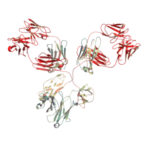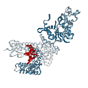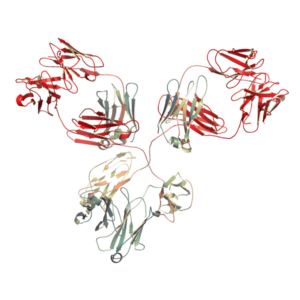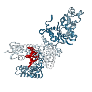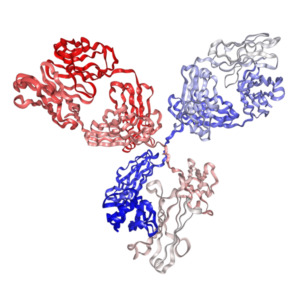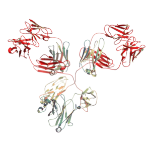
Anti-mouse MHC Class I (H-2Db) Antibody (Clone:28-14-8S) | PA007591.m2a
$150.00 – $900.00
Recombinant mouse IgG2a isotype controls are available. Condition of sample preparation and optimal sample dilution should be determined experimentally by the investigator.
- Details & Specifications
- References
| Catalog No. | PA007591.m2a |
|---|---|
| Product Name | In Vivo Grade Recombinant Anti-mouse MHC Class I (H-2Db) Monoclonal Antibody (Clone: 28-14-8S), Mouse IgG2a Kappa |
| Supplier Name | Syd Labs, Inc. |
| Brand Name | Syd Labs |
| Synonyms | MHC Class I, H-2Db |
| Clone | 28-14-8S. |
| Isotype | Mouse IgG2a Kappa. |
| Source/Host | The anti-mouse MHC Class I (H-2Db) monoclonal antibody (clone: 28-14-8S) was produced in mammalian cells. |
| Specificity/Sensitivity | The in vivo grade recombinant mouse monoclonal antibody (clone: 28-14-8S) specifically binds to mouse MHC Class I (H-2Db). |
| Applications | ELISA, neutralization, functional assays such as bioanalytical PK and ADA assays, and those assays for studying biological pathways affected by the mouse MHC Class I (H-2Db) protein. |
| Form Of Antibody | 0.2 uM filtered solution, pH 7.4, no stabilizers or preservatives. |
| Endotoxin | < 1 EU per 1 mg of the protein by the LAL method. |
| Purity | >95% by SDS-PAGE under reducing conditions and HPLC. |
| Shipping | The in vivo grade recombinant anti-mouse MHC Class I (H-2Db) monoclonal antibody of clone 28-14-8S is shipped with ice pack. Upon receipt, store it immediately at the temperature recommended below. |
| Stability & Storage | Use a manual defrost freezer and avoid repeated freeze-thaw cycles. 12 months from date of receipt, -20 to -70°C as supplied. 1 month from date of receipt, 2 to 8°C as supplied. |
| Note | Recombinant mouse IgG2a isotype controls are available. Condition of sample preparation and optimal sample dilution should be determined experimentally by the investigator. |
| Order Offline | Phone: 1-617-401-8149 Fax: 1-617-606-5022 Email: message@sydlabs.com Or leave a message with a formal purchase order (PO) Or credit card. |
Description
PA007591.m2a: In Vivo Grade Recombinant Anti-mouse MHC Class I (H-2Db) Monoclonal Antibody (Clone: 28-14-8S), Mouse IgG2a Kappa
Recombinant Mouse IgG2a Monoclonal Antibody.
References of Anti-Mouse MHC Class I (H-2Db) Monoclonal Antibody (Clone: 28-14-8S)
1. Identification of immunogenic HLA class I and II neoantigens using surrogate immunopeptidomes.
Tokita, S., et al.Sci Adv. 2024 Sep 20;10(38):eado6491. doi: 10.1126/sciadv.ado6491. PMID: 39292790
“The peptides bound to MHC-I or MHC-II were captured as peptide-MHC complexes using affinity chromatography of W6/32 monoclonal antibody (mAb) for pan HLA-I (HB-95, ATCC), IVA12 mAb for pan HLA-II (HB-145, ATCC), BB7.2 mAb for HLA-A2 (HB-82, ATCC), SF1-1.1.10 mAb for H-2Kd (HB-159, ATCC), 34-5-8S mAb for H-2Dd (HB-102, ATCC), or 28-14-8S mAb for H-2Ld (HB-27, ATCC) coupled to CNBr-activated Sepharose 4B (GE Healthcare) overnight. …In contrast, an in silico prediction algorithm (NetMHCpan4.1) combined with RNA sequencing (RNA-seq) gene expression data predicted 326 candidate neoantigens potentially presented by HLA-A*02:01 from the same mutation data (see Materials and Methods for details). …The in silico–predicted neoantigens consisted of 126 strong binders (%rank NetMHC scores below 0.5) and 200 weak binders (%rank NetMHC scores below 2.0), and we evaluated all 126 strong binders using the tandem IVTT method. …Meanwhile, in silico prediction using NetMHCpan and RNA-seq data predicted 326 HLA-A*02:01 and 312 HLA-I candidate neoantigens (%rank NetMHC scores below 2.0) in CRC135 and UTE003, respectively (Figs. 2A and 3A). …Potential de novo neoantigens were defined as those with wild-type and neoantigen NetMHCpan4.1 scores greater than and less than 2.0, respectively.”
2. Localized ablative immunotherapy drives de novo CD8+ T-cell responses to poorly immunogenic tumors.
Hoover, A. R., et al.Journal for ImmunoTherapy of Cancer. 2022 Oct;10(10):e004973. doi: 10.1136/jitc-2022-004973. PMID: 36253002
“The monoclonal antibodies (clones 28-14-8 (anti-Db) and AF6-88.5.5.3 (anti-Kb)) were used to isolate the MHC class I alleles (Db and Kb) and purify the Db-associated and Kb-associated peptides from the tumors as well as staining of the B16-F10 cells to determine cell surface expression of Db and Kb using flow cytometry. …To determine the expression of the MHC class I molecules (Db and Kb) on the surface of B16-F10 cells, they were seeded at 5×10^5 cells/well in 6-well plates in complete growth medium. …In contrast to B16-F10 tumor cells cultured in vitro which express little-to-no H2-Db or H2-Kb molecules unless treated with IFN-γ (online supplemental figure 2A,B), implanted tumor cells yielded high quantities of MHC class I molecules in vivo. This high expression resulted in the identification of hundreds of H2-Db and H2-Kb tumor-associated peptides. …To identify the TSAs that were exclusively presented by B16-F10 tumor cells and were absent from healthy tissues, the tumor peptidome was searched against a map of murine MHC class I immunopeptidome generated from different tissues of healthy C57BL/6 mice and the peptides expressed on healthy tissues were eliminated. …The anti-Kb monoclonal antibody (clone AF6-88.5.5.3) was purchased from BioXcell. Anti-Db (28-14-8S, ATCC) hybridoma was grown in serum-free media and purified in-house using Protein G Sepharose column (GE Healthcare, Sweden).”
3. Direct Priming of CD8+ T Cells Persists in the Face of Cowpox Virus Inhibitors of Antigen Presentation.
Lin, L. C. W., et al.J Virol. 2021 Apr 26;95(10):e00186-21. doi: 10.1128/JVI.00186-21. PMID: 33692206
“First, degradation of different MHC-I alleles is not the same for US2 and US11; second, US2 can also degrade MHC class II; third, the downregulation of de novo viral antigen presentation on infected cells was not especially strong in these studies. …After clearing the lysate by centrifugation, the peptide-MHC within the supernatant was isolated via immunoaffinity purification using sequential anti-Kb (clone Y-3) and anti-Db (clone 28-14-8S) antibodies cross-linked to protein A Sepharose. …For peptide detection and quantitation, LC-MRM was employed for a panel of known VACV epitopes whose MRM transition parameters have previously been optimized. …Statistical analysis was conducted with GraphPad Prism software or with R. …All cells were maintained in Dulbecco’s modified Eagle medium (DMEM) supplemented with 10% fetal bovine serum (FBS) and l-glutamine.”
4. Identification and Characterization of Rift Valley Fever Virus-Specific T Cells Reveals a Dependence on CD40/CD40L Interactions for Prevention of Encephalitis.
Barbeau, D. J., et al.J Virol. 2021 Nov 9;95(23):e0150621. doi: 10.1128/JVI.01506-21. PMID: 34495703
“Following a 2-day incubation, MHC-bound radioactivity was determined by capturing MHC/peptide complexes on Lumitrac 600 plates (Greiner Bio-One, Frickenhausen, Germany) coated with either Y3 (anti-H-2-Kb), 28-14-8s (anti-H-2-Db, Ld, and Dq), or Y3JP (anti-H-2-IAb,s,u) monoclonal antibodies; bound counts per minute were measured using the TopCount (Packard Instrument Co., Meriden, CT) microscintillation counter. …Research with live virus was conducted at the Center for Vaccine Research’s Regional Biocontainment Laboratory or at the Centers for Disease Control and Prevention (CDC) under biosafety level 3 (BSL3) conditions. …Institutional Animal Care and Use Committee approval was provided by the University of Pittsburgh (protocol no. 17080998 and 19044158) or CDC (protocol no. 2577MCEMOUC or 2629MCEMOUC). …All recombinant virus work was approved by the respective institutional biosafety committees. …Previously, CD4 T cells were identified as a critical component of adaptive immunity that prevented RVFV-mediated encephalitis.”
5. Palladium-Induced Temporal Internalization of MHC Class I Contributes to T Cell-Mediated Antigenicity.
Ito, K., et al.Front Immunol. 2021 Dec 23:12:736936. doi: 10.3389/fimmu.2021.736936. PMID: 35003059
“In this concept, haptenated antigens phagocytosed by APCs are cross-presented by MHC class I after undergoing processing (12). …In other words, the peptide loading onto the MHC class I is derived from haptenated antigens. …In some experiments, prior to co-culture with TG40 cells, peptides on MHC class I were stripped as described previously. …Surface MHC class I expression after PdCl2 treatment. …First, we asked whether Pd-treatment affects MHC class I/peptide complexes. To examine this, mice were injected with ovalbumin (OVA) protein and 12 hours after injection, PdCl2 was injected intraperitoneally.”
6. Identification of Neoantigens in Two Murine Gastric Cancer Cell Lines Leading to the Neoantigen-Based Immunotherapy.
Nagaoka, K., et al.Cancers (Basel). 2021 Dec 27;14(1):106. doi: 10.3390/cancers14010106. PMID: 35008270
“The peptide-MHC class I complexes were captured by affinity chromatography using purified monoclonal antibodies (clone Y-3 for Kb and 28-14-8S for Db) coupled to CNBr-activated Sepharose 4B (GE Healthcare, Chicago, IL, USA). …Peptides bound to MHC class I were eluted with a mild acid (0.2% TFA) and desalted using a Sep-Pak C18 cartridge (Waters Corporation, Milford, MA, USA) with 28% ACN in 0.1% TFA and ZipTip U-C18 (Merck Millipore, Burlington, MA, USA) with 50% ACN in 1% FA. …MHC class I binding peptides were eluted from viable YTN16 cells as described previously. …Neoepitopes derived from the products of these mutated genes were predicted by their estimated binding capacity to MHC class I molecules; 8-, 9-, and 10-mer peptides containing the mutated amino acid were assessed by NetMHCpan and MHCflurry algorithms to predict their binding to and presentation on H-2Db and H-2Kb molecules. …In silico-predicted peptides indeed stabilized MHC class I expression on RMAS cells.”
7. Restoration of cytomegalovirus antigen presentation by gamma interferon combats viral escape.
Hengel, H., et al.J Virol. 1994 Jan;68(1):289-97. doi: 10.1128/JVI.68.1.289-297.1994. PMID: 8254740
“Major histocompatibility complex (MHC) class I molecules bind peptides derived from endogenously synthesized proteins and present them at the cell surface to CD8+ T lymphocytes. …After trypsinization, washing, and counting of cells, peptides were extracted with trifluoroacetic acid from whole cell lysates (~4 x 10^7 cells) and separated first by size on a Sephadex G-25 column and subsequently on a reverse-phase high-pressure liquid chromatography (HPLC) column. …The restoration of antigen presentation therefore comprises two events. …The following monoclonal antibodies (MAbs) were used: 28-14-8S (anti-Ld) (ATCC HB 27), 64-3-7 (anti-Ld, selective for Ldalt), 30-5-7 (anti-Ld, selective for β2m-associated Ld), 34-5-8S (anti-Dq) (ATCC HB 102), GK1.5 (anti-CD4) (ATCC TIB 207), YTS-169.4 (anti-CD8), YTS-191.1 (anti-CD4+), R4-6A2 (anti-murine IFN-γ) (ATCC HB 170), AN-18 (anti-murine IFN-γ), and V1q (anti-murine tumor necrosis factor). Anti-MCMV serum was collected from latently infected BALB/c mice. …In brief, quantitative precipitations were performed sequentially with ascitic fluids of the MAb 64-3-7, followed by 28-14-8S and 34-5-8S.”
For more technical references or data sheets regarding the Recombinant Anti-Mouse MHC Class I (H-2Db) Monoclonal Antibody (Clone: 28-14-8S) formats, please contact our scientific support team at message@sydlabs.com.
In Vivo Grade Recombinant Anti-mouse MHC Class I (H-2Db) Monoclonal Antibody (Clone: 28-14-8S), Mouse IgG2a Kappa from: In Vivo Grade Recombinant Anti-mouse MHC Class I (H-2Db) Monoclonal Antibody (Clone: 28-14-8S), Mouse IgG2a Kappa: PA007591.m2a
You may also like…
-
 Select optionsQuick View