

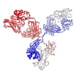
Anti-Digoxigenin Monoclonal Antibody (Clone: 5A7) | PA007704.m1
$150.00 – $300.00
Recombinant mouse IgG1 isotype controls are available. Condition of sample preparation and optimal sample dilution should be determined experimentally by the investigator.

- Details & Specifications
- References
| Catalog No. | PA007704.m1 |
|---|---|
| Name | Anti-Digoxigenin Monoclonal Antibody (Clone: 5A7) | PA007704.m1 |
| Product Name | Recombinant Anti-Digoxigenin Monoclonal Antibody (Clone: 5A7), Mouse IgG1 Kappa |
| Supplier Name | Syd Labs, Inc. |
| Brand Name | Syd Labs |
| Synonyms | Anti-digoxin Antibody, Anti-DIG Antibody |
| Clone | 5A7. |
| Isotype | Mouse IgG1 kappa. |
| Source/Host | The recombinant anti-Digoxigenin monoclonal antibody (clone: 5A7) was produced in mammalian cells. |
| Specificity/Sensitivity | The recombinant anti-Digoxigenin monoclonal antibody (clone: 5A7) specifically binds to Digoxigenin. |
| Applications | Western Blot, ELISA, Immunohistochemistry (IHC). |
| Form Of Antibody | 0.2 μM filtered solution of 1x PBS. |
| Purity | >95% by SDS-PAGE under reducing conditions and HPLC. |
| Shipping | The recombinant anti-Digoxigenin monoclonal antibody of clone 5A7 is shipped with ice pack. Upon receipt, store it immediately at the temperature recommended below. |
| Stability & Storage | Use a manual defrost freezer and avoid repeated freeze-thaw cycles. 12 months from date of receipt, -20 to -70°C as supplied. 1 month from date of receipt, 2 to 8°C as supplied. |
| Note | Recombinant mouse IgG1 isotype controls are available. Condition of sample preparation and optimal sample dilution should be determined experimentally by the investigator. |
| Order Offline | Phone: 1-617-401-8149 Fax: 1-617-606-5022 Email: message@sydlabs.com Or leave a message with a formal purchase order (PO) Or credit card. |
Description
PA007704.m1: Recombinant Anti-Digoxigenin Monoclonal Antibody (Clone: 5A7), Mouse IgG1 Kappa
Recombinant mouse IgG1 monoclonal antibody.
Background of Anti-digoxigenin Monoclonal Antibody
Recombinant mouse anti-digoxigenin antibodies recognize digoxigenin (digoxin, DIG) attached to proteins, peptides, nucleic acids, or solid matrices, and can be offered in fluorophore and enzyme-conjugated versions.
The anti-digoxigenin antibody (anti-DIG antibody) can be used as a second-step reagent in various applications for detecting digoxigenin-conjugated targets. The digoxigenin-anti-digoxigenin system is widely used in immunoassay signal amplification techniques because an anti-digoxigenin antibody may bind digoxigenin with high affinity.
Please contact us to ask for a quote for the engineered anti-digoxigenin antibody mutants of various isotypes, and mutants with Avi-, His-, and DYKDDDDK-tags, and biotinylated antibodies and mutants. A variety of conjugates (such as dyes and fluorophores) with the anti-digoxigenin antibodies and mutants are available.
References of Anti-Digoxigenin Antibody (Clone: 5A7)
1. Complexation of drug and hapten-conjugated aptamer with universal hapten antibody for pancreatic cancer treatment
Sun Il Choi, et al.J Control Release. 2023 Aug;360:940-952. doi: 10.1016/j.jconrel.2023.03.048. PMID: 37001565
“Monomethyl auristatin E was used as a cytotoxic drug, digoxigenin acted as a hapten, and the humanized anti-digoxigenin antibody served as a universal carrier of the aptamer. Additionally, we generated the PAp7T8-DOligobody, which is coupled MMAE and the anti-digoxigenin (DIG) antibody, a novel humanized universal antibody. Owing to a lack of reliable markers and therapeutic targets, pancreatic ductal adenocarcinoma (PDAC) remains the most lethal malignant tumor despite numerous therapeutic advances. In this study, we utilized cell-SELEX to isolate a DNA aptamer recognizing the natural conformation of the target on the cell surface. Therefore, PAp7T8-DOligobody represents a promising novel therapeutic delivery platform for PDAC.”
2. Nanoparticle-Based Visual Detection of Amplified DNA for Diagnosis of Hepatitis C Virus
Soo-Kyung Kim, et al.Biosensors (Basel). 2022 Sep 9;12(9):744. doi: 10.3390/bios12090744. PMID: 36140129
“A mixture of 2 μL of streptavidin-coated magnetic bead suspension (1 μm, 10 mg/mL Dynabears MyOne Streptavidin C1; Life Technologies, Grand Island, NY, USA) and 1 μL of anti-digoxigenin antibody-coated polystyrene particles (250–350 nm, 5 mg/mL anti-digoxigenin alpha-donor beads in PBS pH 7.2; PerkinElmer, Waltham, MA, USA) was added to PCR tubes containing amplified DNA. Among commercially available anti-digoxigenin antibody-coated particles, we selected blue polystyrene particles (250–350 nm anti-digoxigenin alpha-donor beads, PerkinElmer; 5 mg/mL) to capture the digoxigenin-labeled reverse primer. Our STAT-DNA test can be performed with a broad range of nano- (250–300 nm-sized anti-digoxigenin antibody-coated polystyrene particles) to micro-sized particles (1 μm-sized streptavidin-coated magnetic particles). The streptavidin-coated magnetic particle–DNA–alpha particle complex (C) showed the most prominent aggregation. Among the commercially available anti-digoxigenin antibody-coated particles, the alpha-donor particles successfully produced the aggregation of DNA amplicons with streptavidin-coated microparticles.”
3. Target-responsive triplex aptamer nanoswitch enables label-free and ultrasensitive detection of antibody in human serum via lighting-up RNA aptamer transcriptions
Jirong Yang, et al.Talanta. 2024 Oct 1;278:126455. doi: 10.1016/j.talanta.2024.126455. PMID: 38917548
“Accurate and sensitive monitoring of the concentration change of anti-digoxigenin (Anti-Dig) antibody is of great importance for diagnosing infectious and immunological diseases. In this study, we used anti-digoxigenin (Anti-Dig) antibody as a detection model and devised a stable triplex aptamer nanoswitch coupled with Taq DNA polymerase through Watson-Crick and Hoogsteen base pairings. At present, various RNA aptamers such as Spinach, Broccoli, Pepper, Mango, and Malachite Green (MG) have been isolated. To meet these challenges, we herein report a novel label-free and ultrasensitive fluorescence turn-on antibody detection assay. Compare with existing methods, our method exhibits three outstanding advantages.”
4. Fluorescent In Situ Hybridization in Embryos of the African Turquoise Killifish Nothobranchius furzeri
Philip B Abitua.Cold Spring Harb Protoc. 2024 Feb 1;2024(2):107805. doi: 10.1101/pdb.prot107805. PMID: 36922000
“Anti-digoxigenin antibodies conjugated to horseradish peroxidase (POD) are then bound to the probe and reacted with fluorescently labeled tyramide. Precisely where and when a given gene is expressed is crucial for our understanding of developmental and cell biology but determining this is often constrained by detection limits. Here, we describe a technique for visualization of low-copy mRNA in Nothobranchius furzeri embryos using tyramide signal amplification (TSA). In this protocol, an anti-sense digoxigenin-labeled RNA probe is hybridized to mRNA in situ. Combining this method with a counterstain, such as DAPI, allows for the detection of mRNA at a single-cell resolution.”
5. Efficient Cross-Screening and Characterization of Monoclonal Antibodies against Marek’s Disease Specific Meq Oncoprotein Using CRISPR/Cas9-Gene-Edited Viruses
Man Teng, et al.Viruses. 2023 Mar 23;15(4):817. doi: 10.3390/v15040817. PMID: 37112797
“Marek’s disease (MD) caused by pathogenic Marek’s disease virus type 1 (MDV-1) is one of the most important neoplastic diseases of poultry. Confocal microscopic analysis of cells stained with these antibodies confirmed the nuclear localization of Meq in MDV-infected CEF cells and MDV-transformed MSB-1 cells. Amino acid variations of Meq do exist among different MDV-1 epidemic strains, even among different strains within the same pathotypes that show genetic diversity and varying virulence. For a long time, the molecular mechanism of MD tumorigenesis induced by Meq has been an attractive research topic, hence monoclonal antibodies (mAbs) against Meq are becoming crucial. As expected, our data exemplified a new strategy for the efficient screening and characterization of specific mAbs against MDV-1 Meq proteins, providing a new approach for the future generation of other virus-specific mAbs.”
Syd Labs provides the following in vivo grade recombinant anti-Biotin antibodies:
In Vivo Grade Recombinant Anti-Biotin Antibodies (Clone F1)
Syd Labs provides the following recombinant anti-Fluorescein/Digoxin/Biotin/Phospho-Tyrosine monoclonal antibodies:
Recombinant Anti-Fluorescein Monoclonal Antibodies (Clone 4-4-20 (enhanced))
Recombinant Anti-Fluorescein Monoclonal Antibodies (Clone 4M5.3)
Recombinant Anti-Digoxin Monoclonal Antibodies (Clone 5A7)
Recombinant Anti-Digoxin Monoclonal Antibodies (Clone 26-10)
Recombinant Anti-Biotin Monoclonal Antibodies (Clone F1)
Recombinant Anti-Biotin Monoclonal Antibodies (Clone M33)
Recombinant Anti-Phospho-Tyrosine Monoclonal Antibodies (Clone Py20)
Recombinant Anti-Phospho-Tyrosine Monoclonal Antibodies (Clone 4G10)
Recombinant Anti-Digoxigenin Monoclonal Antibody (Clone: 5A7), Mouse IgG1 Kappa from: Recombinant Anti-Digoxigenin Monoclonal Antibody (Clone: 5A7), Mouse IgG1 Kappa: PA007704.m1 Syd Labs
You may also like…
- Select optionsQuick View
- Select optionsQuick View
- Select optionsQuick View
- Select optionsQuick View
- Select optionsQuick View
- Select optionsQuick View
- Select optionsQuick View
- Select optionsQuick View