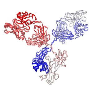

Anti-mouse CD8a Antibody for Flow Cytometry | PA007499.h1Fs


$45.00 – $90.00
Recombinant rat IgG2a isotype controls and Recombinant human IgG1 isotype controls are available. Condition of sample preparation and optimal sample dilution should be determined experimentally by the investigator.
- Details & Specifications
- References
| Catalog No. | PA007499.h1Fs |
|---|---|
| Product Name | Recombinant Anti-mouse CD8b Monoclonal Antibody (Clone: YTS 156.7), Human IgG1 Fc Silent Kappa |
| Data Sheet | 📄 Download Product Data Sheet (PDF) |
| Supplier Name | Syd Labs, Inc. |
| Brand Name | Syd Labs |
| Synonyms | CD8 alpha, T8, Lyt2, Ly-2, T-cell surface glycoprotein CD8 alpha chain, CD_antigen CD8a |
| Clone | YTS 105.18. |
| Isotype | Human IgG1 Fc Silent Kappa |
| Source/Host | The anti-mouse CD8a monoclonal antibody (clone: YTS 105.18) for Flow Cytometry was produced in mammalian cells. |
| Specificity/Sensitivity | The recombinant YTS 105.18 antibody for Flow Cytometry specifically binds to mouse CD8a protein. |
| Applications | Flow cytometry (FC), and Immunohistochemistry - Frozen (IHC-F). |
| Form Of Antibody | 0.2 uM filtered solution, pH 7.4, containing 0.09% sodium azide. |
| Conjugate | Non-conjugated. |
| Purity | >95% by SDS-PAGE under reducing conditions. |
| Shipping | The Recombinant Anti-mouse CD8a Monoclonal Antibody (Clone: YTS 105.18) for Flow Cytometry, Human IgG1 Fc Silent Kappa is shipped with ice pack. Upon receipt, store it immediately at the temperature recommended below. |
| Stability & Storage | Use a manual defrost freezer and avoid repeated freeze-thaw cycles. 12 months from date of receipt, 2 to 8°C as supplied. Do not freeze it. |
| Note | Recombinant human IgG1 isotype controls are available. Condition of sample preparation and optimal sample dilution should be determined experimentally by the investigator. |
| Order Offline | Phone: 1-617-401-8149 Fax: 1-617-606-5022 Email: message@sydlabs.com Or leave a message with a formal purchase order (PO) Or credit card. |
Description
PA007499.h1Fs: Recombinant Anti-mouse CD8a Monoclonal Antibody (Clone: YTS 105.18) for Flow Cytometry, Human IgG1 Fc Silent Kappa
The rat anti-mouse CD8a monoclonal antibody YTS 105.18 (Rat IgG2a kappa) reacts with the mouse CD8a protein (T-cell surface glycoprotein CD8 alpha chain) encoded by the mouse CD8A gene that encodes the CD8a chain of the dimeric CD8 protein. The mouse CD8 protein is primarily responsible for cell-mediated immune defense and T-cell development. CD8A has been widely reported as a potential prognosis and diagnostic marker for several diseases, such as inflammatory disorders and tumors. It was shown that the anti-mouse CD8a rat monoclonal antibody (clone number: YTS 105.18) can be used for depleting activity in vivo.
Our recombinant YTS 105.18 antibodies have a part (variable regions) or complete amino acid sequences of the rat anti-mouse CD8a monoclonal antibody (hybridoma clone name or number: YTS 105.18).
References of Anti-Mouse CD8b Monoclonal Antibody (Clone: YTS 156.7)
1. Adding liposomal doxorubicin enhances the abscopal effect induced by radiation/αPD1 therapy depending on tumor cell mitochondrial DNA and cGAS/STING.
Wang L, et al.J Immunother Cancer. 2023 Aug;11(8):e006235. PMID: 37640480
“In some experiments, CD8+ T cells were depleted by injecting 200 µg/mouse of anti-CD8α antibodies (clone 2.43, ATCC) or anti-CD8β (clone YTS 156.7.7, ECACC European collection of cell cultures) antibodies i.p. 2 days before hRT, on the day of the first hRT, and once weekly thereafter. Flow-cytometric analyses of tumor-infiltrating lymphocytes (TILs) and single-cell suspensions from tumors, blood, and lymph nodes were performed using the following antibodies/MHC tetramers. M8 tetramer-PE (H-2Kb, p15E) with CD8-AF700 (clone KT15/MBL) were used to detect tumor-specific CD8+ T cells. CXCR3-APC-Fire750 (clone CXCR3-173) was from Miltenyi Biotec. MHCII-AF700 (clone M5/114.15.2), CD45-BV510 (clone 30-F11), CD3-PE-Cy5.5 or -FITC (clone 145-2C11), CD49b-FITC (clone DX5), CD19-FITC (clone 1D3), F4/80-FITC (clone 1BM8), CD8-APC (clone 53-6.7), Tim-3-BV605 (clone RMT3-23), Ki-67-BV605 (clone 16A8), CD11c-BV650 (clone N418), CD11b-PE-Cy7 (clone M1/70), CD86-BV421 (clone GL1), Ly6C-PerCP-Cy5.5 (clone HK1.4) and CD103-PE-CF594 (clone M290) were from…”
2. CD8+ T Cells Regulate Monopoiesis and Circulating Ly6C-high Monocyte Levels in Atherosclerosis in Mice.
Cochain C, et al.Circ Res. 2015 Jul 17;117(3):244-53. PMID: 25991812
“Eight- to 10-week-old male mice were placed on an atherogenic diet (15% milk fat, 1.25% cholesterol; Altromin) for 6 weeks, and treated with weekly intraperitoneal injections of 250 µg of rat antimouse-CD8α (clone YTS-169, referred to as anti-CD8α or a-CD8α), rat antimouse-CD8β (clone YTS-156.7.7, referred to as anti-CD8β or a-CD8β), or isotype-matched irrelevant rat-anti-Phyt1 (clone AFRC-MAC51, referred to as control). For in vivo IFN-γ-neutralization, mice received intraperitoneal injections of 250 µg of rat antimouse IFN-γ antibody (clone XMG1.2) or irrelevant isotype-matched rat anti–β-galactosidase antibody (clone GL113) every 48 hours for 3 weeks. Intracellular labeling of TNF-α, IL-12, IL-17A, and IFN-γ was performed after PMA (phorbol-12-myristat-13-acetat)/ionomycine/brefeldin stimulation for 4 hours using the Becton Dickinson Cytofix/Cytoperm Kit. Bone marrow–derived macrophages or SV40-transformed mouse endothelial cells (SVEC) were serum starved and cultured alone or with isolated CD8+ T cells at a 1:1 ratio in RPMI containing 0.5% FCS, 5 µg/mL anti-CD3, and 1 µg anti-CD28 antibody, with the addition of 10 µg/mL anti–IFN-γ-neutralizing antibody or 10 µg/mL of isotype control antibody. Chemokine (CC-motif) ligand 2 (CCL2), chemokine (CXC-motif) ligand 1 (CXCL1), and CCL5 levels in serum and cell culture supernatants were measured using the corresponding ELISA Development Kits according to the manufacturer’s instructions (Peprotech).”
3. Effects of T-lymphocyte depletion on muscle fibrosis in the mdx mouse.
Morrison J, et al.Am J Pathol. 2005 Jun;166(6):1701-10. PMID: 15920155
“Seven and nine days after the thymectomy, each mouse received intraperitoneal injections of CD4 (YTS 191.1.2 and YTA 3.1.2) or CD8 (YTS 156.7.7 and YTS 169.4.7) antibodies or both together. To investigate the role of T lymphocytes in fibrosis, we depleted either CD4 or CD8 or both subsets of T lymphocytes from 4-week-old mdx mice by use of thymectomy followed by specific antibody administration 7 and 9 days after thymectomy. Mice that had less than 2.5% circulating or splenic CD4 cells were grouped together and represent the highest CD4-depleted animals, which was equivalent to more than 97.5% depletion. Overall results showed that hydroxyproline levels in the diaphragm of CD4, CD8, or CD4/8 double-depleted mdx mice were indistinguishable from those seen in untreated mdx. In both single- and double-depleted mdx and C57BL/10 mice, some T lymphocytes were still present within the diaphragm.”
4. Insulin immunization of nonobese diabetic mice induces a protective insulitis characterized by diminished intraislet interferon-gamma transcription.
Muir A, et al.J Clin Invest. 1995 Feb;95(2):628-34. PMID: 7860747
“Synergistic depleting monoclonal antibodies against CD4+ (YTS 191.1 and YTA 3.1.2) and CD8+ (YTS 169.4 and YTS 156.7) T lymphocytes were kindly provided by Dr. Anne Cooke (Cambridge University, UK). Control mice received injections of either PBS or an irrelevant IgG monoclonal antibody against influenza A nucleoprotein (kindly provided by Dr. Parker Small, University of Florida, Gainesville). Starting 1 wk after completing a two-dose immunizing course of B-chain insulin at 4 and 8 wk of age, one intravenous dose followed by two intraperitoneal monoclonal antibody or control treatments were administered. We performed flow cytometric analyses of splenocytes from treated or control mice on a FACScan (Becton-Dickinson, San Jose, CA) after staining with GK1.5 (anti-CD4) and clone 53-6.7 (anti-CD8) antibodies (Pharmingen, San Diego, CA). Before transfer, six donors received treatments with anti-CD4 antibodies, eight animals were treated with anti-CD8 antibodies, and seven received injections of either PBS (n = 3) or the irrelevant IgG monoclonal antibody (n = 4).”
5. Induction of classical transplantation tolerance in the adult.
Qin SX, et al.J Exp Med. 1989 Mar 1;169(3):779-94. PMID: 2647894
“Of the two CD8 mAbs, YTS 169.4 is anti-Lyt-2 and YTS 156.7 (anti-mouse CD8b antibody) is anti-Lyt3. The two pairs of synergistic mAbs used in the experiments were all of rat IgG (rIgG)2b isotype. All antibodies were obtained from (DA x LOU)F1 rat ascitic fluid, purified by ammonium sulfate precipitation and dialyzed into PBS. Preparation of mAb F(ab’)2 Fragments. F(ab’)2 fragments of YTS 191.1 and YTS 169.4 were made by the method of Rousseaux et al. The purity of F(ab’)2 products was confirmed by SDS-PAGE and by complete inhibition of in vitro complement lysis by parental antibodies as reported elsewhere.”
For more technical references or data sheets regarding the Recombinant Anti-Mouse CD8b Monoclonal Antibody (Clone: YTS 156.7) formats, please contact our scientific support team at message@sydlabs.com.
Syd Labs provides the following recombinant anti-human CD8a monoclonal antibodies:
Recombinant anti-human CD8a antibodies (clone OKT8),In vivo grade
Recombinant anti-human CD8a antibodies (clone OKT8) for flow cytometry
Syd Labs provides the following recombinant anti-mouse CD8a monoclonal antibodies:
Recombinant anti-mouse CD8a antibodies (clone 2.43), In vivo grade
Recombinant anti-mouse CD8a antibodies (clone YTS 169.4)R, In vivo grade
Recombinant anti-mouse CD8a antibodies (clone YTS 105.18), In vivo grade
Syd Labs provides the following recombinant anti-mouse CD8b monoclonal antibodies:
Recombinant anti-mouse CD8b antibodies (clone YTS 156.7), In vivo grade
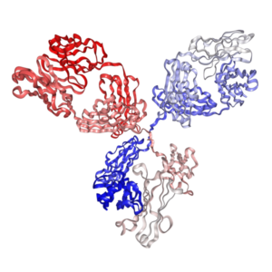
Anti-mouse CD8a Antibody (YTS 105.18) for Flow Cytometry from: Recombinant Anti-mouse CD8a Monoclonal Antibody (Clone: YTS 105.18), Human IgG1 Fc Silent Kappa: PA007499.h1Fs Syd Labs
You may also like…
-
 Select optionsQuick View
-
 Select optionsQuick View
-
 Select optionsQuick View
-
 Select optionsQuick View